Download

1 / 36
400 likes | 1.87k Vues
1. FES FOR THE PAINFUL HEMIPLEGIC SHOULDER. Overview Summary of research evidence Implications for PT practice. Arlene Mendoza, Janet Sanabria, Telan Nelson, Amanda Lonsdale, Allison Pieracci, & Heather Nordberg . Learning Objectives.

E N D
FES FOR THE PAINFUL HEMIPLEGIC SHOULDER Overview Summary of research evidence Implications for PT practice Arlene Mendoza, Janet Sanabria, Telan Nelson, Amanda Lonsdale, Allison Pieracci, & Heather Nordberg
Learning Objectives By the end of the presentation the learner will be able to: • Describe the incidence and etiology of hemiplegic shoulder pain (HSP). • Explain the methods for preventing and treating HSP. • Define Functional Electrical Stimulation (FES). • Explain how FES possibly decreases HSP & increases function. • Conclude whether the use of FES post-stroke to treat HSP is effective and recommended. • Explain how time post stroke relates to effectiveness of FES treatment.
Review of the Hemiplegic Shoulder • Muscle tone change in the UE from the early to late phase of recovery: • Continuum from flacidity-> hypotonicity-> normal tone-> hypertonicity-> rigidity • Problems that can result from flaccidity: • GH subluxation and stretching of the capsule, ligaments, muscles, and nerves • Problems that can result from hypertonicity: • Flexion synergy pattern in which shoulder is add and IR. Teasell, R. et al. (2008)
Incidence of HSP • In a sample of 10 studies, the incidence of HSP varied from 9-73% of hemiplegic stroke patients. • The onset of HSP ranged from 2 weeks to 6 months post-stroke. • There is no standardized method for determining/measuring the epidemiology of HSP. • The incidence of HSP seems to increase over time following stroke. • Teasell, R. et al. (2008)
Etiology of HSP • Multi-factorial • GH subluxation • Impingement • Decreased ROM/Frozen Shoulder • CRPS (Complex Regional Pain Syndrome) • Spasticity Ada, L., & Foongchomcheay, A (2002), Chantraine, A. et al. (1999) Teasell, R. et al. (2008), Walsh, K. (2001), Vaugnat, H. et al. (2003).
Prevention and Treatment of HSP • Acute: • Prevention and Treatment: • Positioning/PROM/Handling/Family education/E-stim • Chronic: • Treatment: • Positioning • Injections (steroid, Botox) • Aromatherapy and acupressure • Electrical stimulation • TENS • FES Teasell, R. et al. (2008), Walsh, K. (2001), Vaugnat, H. et al. (2003)
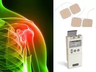
Definition of FES “FES utilizes electrical currents to activate nerves in areas of a patient’s body affected by paralysis, stroke, traumatic brain injuries, and other neurological disorders to restore some movement & function.” - Cleveland FES Center, OH
How Does FES Reduce HSP? Shoulder subluxation and ER One proposed mechanism of reducing HSP, however it is uncertain that shoulder subluxation is the cause of HSP. Chantraine, A. et al. (1999), Ada, L., & Foongchomcheay (2002) , Wang, RY et al. (2000), Price, C & Pandyan, A (2009), Teasell, R. et al. (2008)
How Does FES Reduce HSP?(continued) Other proposed mechanisms include: • Stimulation of somatosensory cortex by augmented sensory feedback • Increased proprioceptive stimulation • Repetitive movements important for motor re-learning • Increased muscle strength • sNMESof cutaneous sensory nerves may modulate pain via gating pathways and central neuromodulation. Church, C. et al. (2006)
Shoulder Pain and Dysfunction in Hemiplegia: Effects of Functional Electrical Stimulation.Chantraine, A. et al. (1999) • Controlled study of 24 months beginning in the first month after onset of stroke. • Included CVA and Brain Injury subjects. • 120 patients with a subluxed and painful hemiplegic shoulder (HSP). • Patients were assigned to a control group or treatment group for a total of 5 weeks • Control Group: conventional therapy (60 subjects) • Treatment Group: FES and conventional therapy (60 subjects)
Treatment group: FES with Conventional TherapyChantraine, A. et al. (1999) • Sequence of FES Program • 1st Sequence • 90 min, rectangular biphasic, 8 Hz, 350 usec, 1:5, 4 channel • 2nd Sequence • 30 min, 40 Hz • 3rd Sequence • 10 min, 1 Hz
Reduction in PAIN Treatment group vs. Control groupChantraine, A. et al. (1999)
Improvement in Subluxation GradesTreatment group vs. Control groupChantraine, A. et al. (1999) de Bats Subluxation Scale Grade 1: Widening of the GH joint line or outward gliding of humeral head. No rupture of the scapulohumeral girdle. Grade 2: Evidence of the beginning of scapulohumeral girdle rupture. Grade 3: The scapulohumeral girdle rupture is complete. The joint is somewhat impaired.
Improvement in Recovery of Motor FunctionTreatment group vs. Control groupChantraine, A. et al. (1999)
ResultsChantraine, A. et al. (1999) • At all measurement stages the FES group had statistically significant improvements in subluxation, pain, and ROM as compared to the control group. Maintained for at least 24 mo’s. • Treatment group: Maximum improvement in pain, subluxation, and motor recovery was observed at 6 months. • Control group: Slow & progressive improvement reaching a max improvement after 1 yr. • Overall: Two thirds of the cases improved in pain, subluxation, and remained constant up to 24 months.
ConclusionChantraine, A. et al. (1999) FES appears to decrease pain, subluxation, improve ROM, and increase motor function and therefore directly influences the degree and rate of recovery.
Intramuscular Electrical Stimulation for Hemiplegic Shoulder Pain Yu DT, Chae, J. et al (2004) • Subjects: 61 • Treatment Group: 32 (intramuscular NMES) • Control Group: 29 (Cuff-type sling) • Inclusion criteria: • >12 weeks post-stroke • Pain rating >2 on the 11 pt. NRS • 1/2 fingerbreadth of inferior glenohumeral subluxation
Study Parameters Yu DT, Chae, J. et al (2004) • Treatment Group: • Intramuscular stimulation 6 hours/day for 6 weeks • 20 sec on time/10 sec off time • 20 mA and 10 - 200㎲ • Intramuscular electrodes placed in the supraspinatus, posterior deltoid, middle deltoid, and upper trapezius. • The electrodes were placed in the clinic and left in for the duration of the study. • Control Group: • Cuff-type hemisling for 6 weeks
Outcome Measures Yu DT, Chae, J. et al (2004) • Primary Outcome Measures: • BPI 12--A pain questionnaire that assesses pain intensity (0-10) scale as well as interference of pain in daily activites. • Secondary Outcome Measures: • BPI question 23 • Subluxation (assessed radiographically) • Pain- free passive ER ROM • Hemiparetic upper limb strength and coordination measured through the Fugl-Meyer motor assessment • Spasticity assessed with Ashworth scale • Upper limb-related activity limitation assessed by FIM instrument and Arm Motor Ability Test
Results: Pain improvement Early group vs. Late group Chae, J. et al (2007) Percent of treatment successes based on the 30% success criterion. ES = electrical stimulation. *P = .001. **P < .001. Percent of treatment successes based on the 2- point success criterion. ES = electrical stimulation. *P = .001. **P < .001.
Results: Early Group vs. Late GroupMean change in BPI 12 scoresChae, J. et al (2007) Early group (<77 weeks post stroke) Late group (>77 weeks post stroke)
Results: Treatment Group vs. Control GroupChae, J. et al (2007) • At end of treatment (EOT), 84% of the ES group experienced a ≥2 pain scale reduction compared to 31% of the control group. • At 12 months, 78% of the ES group experienced a ≥2 pain scale reduction compared to 52% of the control group. • A significantly higher success rate was seen for the ES group compared to the control group at EOT but not at 3, 6, and 12 months. • There was no significant difference in any of the secondary outcomes measured.
Conclusion Chae, J. et al (2007) • FES can be beneficial for HSP if treated early (<77 weeks), and effects can be seen up to 12 months after treatment. • Late treatment (>77 weeks) showed no significant improvements and any effects are only seen short-term. • The treatment group had a higher success rate at EOT, but there was no significant difference between the treatment group and the control group at all of the follow-up measurements.
RCT to Evaluate the Effect of sNMES to the Shoulder After Acute StrokeChurch, C. et al. (2006) • 176 Stroke patients, within 10 days post stroke • Treatment Group: • (90 patients) surface neuromuscular electric stimulation (sNMES) and stroke unit rehab • One electrode over supraspinatus and one over posterior deltoid • 30 Hz; 15sec on/ 15sec off (3 sec ramp) • Increase intensity until visible contraction • Treated 1hr, 3x/day for 1 month • Control Group: • (86 patients) “sham” sNMES and stroke unit rehab
Outcome MeasuresChurch et al. (2006) • Primary (at 3 months): • Action Research Arm Test (ARAT) • Secondary (at 4 weeks and at 3 months): • Motricity Index • Frenchay Arm Test • 0-10 Numerical Pain Rating Scale (UE) • 5-point adjectival scale (UE pain) • Star Cancellation (for cortical function) • Participants’ views regarding the sNMES • Global health status at 3 months (Nottingham Health Profile and Oxford Handicap Scale)
ResultsChurch et al. (2006) • Results at 4 weeks: • There were no significant differences in any of the outcome measures between the control group and the treatment group. • Results at 3 months: • There were no statistically significant differences in: • Arm function (ARAT total) • Upper limb pain • Star Cancellation • Global health status
Is sNMEShazardous for stroke patients with severe upper limb impairment?Church et al. (2006) There was a statistically significant difference in the grasp and gross subsections of the ARAT and Frenchay Arm Test and the arm portion of the Motricity Index in favor of the control group.
Many hypotheses why poor outcome with severely impaired UEChurch et al. (2006) • Abnormal afferent stimulation causes maladaptive plasticity. • Early over-use of the affected arm • Unable to report adverse events or wrong delivery • Overstimulation leading to shoulder subluxation • May have promoted learned non-use of this arm.
ConclusionChurch et al. (2006) For typical stroke patients treated in stroke rehab units: • sNMESdoes NOT improve upper limb function, nor decrease pain after acute stroke. • Routine use of sNMESCANNOT be recommended as it can pose potential negative consequences in those with initial severe impairment. • Further research is needed to determine if there is a benefit to using sNMES for specific patient populations.
The Evidence-Based Review of Stroke RehabilitationTeasell, R. et al., 2008 • Canadian Systematic Review • Over 15 Studies included • There is conflicting evidence that FES reduces pain, improves function and reduces subluxation after stroke. • FES may not help with recovery of hemiplegic shoulder. www.ebrsr.com
Implications for PT • Standardization is needed in reporting HSP • Inconclusive if FES reduces HSP • Inconclusive if FES improves function • FES may be beneficial for treating GH subluxation • Earlier initiation of FES treatment may result in a better outcome
Review Learning Objectives Describe the incidence and etiology of hemiplegic shoulder pain (HSP). Explain the methods for preventing and treating HSP. Define Functional Electrical Stimulation (FES). Explain how FES possibly decreases HSP & increases function. Conclude whether the use of FES post-stroke to treat HSP is effective and recommended. Explain how time post stroke relates to effectiveness of FES treatment.
REFERENCES • Ada, L., & Foongchomcheay, A. (2002). Efficacy of electrical stimulation in preventing or reducing subluxation of the shoulder after stroke: A meta-analysis. Australian Journal of Physiotherapy, 48: 257- 267. • Chantraine, A., Baribault, A., Uebelhart, D., & Gremion, G. (1999). Shoulder pain and dysfunction in hemiplegia: Effects of functional electrical stimulation. Archives of Physical Medicine and Rehabilitation, 80: 328-331. • Chae J et al. Intramuscular Electrical Stimulation for Hemiplegic Shoulder Pain: a 12 month follow up of a multiple center, randomized clinical trial. Am J Phys Med Rehabil. 2005;84:832-842. • Church, C., Price, C., Pandyan, AD., Huntley, S., Curless, R., Rodgers, H. (2006). Randomized controlled trial to evaluate the effect of surface neuromuscular electrical stimulation to the shoulder after acute stroke. Stroke, 37: 2995–3001.
REFERENCES Price, CIM., & Pandyan, AD. (2009). Electrical stimulation for preventing and treating post-stroke shoulder pain. Cochrane Database of Systematic Reviews 2009 (1). Scott, Tom. "Functional Electrical Stimulation: The Future of Rehabilitation." Cleveland FES Center. United Spinal’s Action Online Magazine, 17 Nov. 2008. Web. 24 Apr. 2010. <http://fescenter.org/index.php?view=article&id=98:functional-electrical-stimulation-the-future-of-rehabilitation&option=com_content&Itemid=15> Teasell, R., Foley, N., Bhogal, S. (2008). Version 11: Painful hemiplegic shoulder. Obtained from the WWW April 7, 2009 at http://www.ebrsr.com/reviews_details.php?16 Vaugnat, H. & Chantraine, A. (2003). Shoulder pain in hemiplegia revisited:Contribution of functional electrical stimulation and other therapies. Journal of Rehabilitative Medicine, 35: 49-56.
REFERENCES • Wang, RY., Chan, RC., & Tsai, MW. (2000). Functional electrical stimulation on chronic and acute hemiplegic shoulder subluxation. American Journal of Physical Medicine and Rehabilitation, 79 (4): 385-390. • Walsh, K. (2001). Management of shoulder pain in patients with stroke. Postgraduate Medical Journal, 77: 645-649. • Yu DT, Chae J, Walker ME, et al. Intramuscular neuromuscular electrical stimulation for post-stroke shoulder pain: a multi-center randomized clinical trial. Arch Phys Med Rehabil. 2004;85:695-704.