Download
1 / 39

E N D
Chromosomes Dr. A. A. Abougabal
Chromosomes • In eukaryotic cells the genetic material is organized into a complex structure composed of DNA and proteins and localized in a specialized compartment, the nucleus. This structure was calledchromatin (from the Greek "khroma" meaning coloured and "soma" meaning body). Close to two meters of DNA in each cell must be assembled into a small nucleus of somemm in diameter. Despite this enormous degree of compaction, DNA must be rapidly accessible to permit its interaction with protein machineries that regulate the functions of chromatin: • · replication, • · repair and • · recombination.
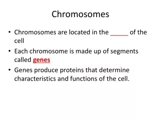
Chromosomes • Chromosomes are also located in the nucleus. When a cell is "resting" i.e. in the interphase not dividing, • Chromosomes are also located in the nucleus. • When a cell is "resting" i.e. not dividing, the chromosomes are organized into long entangled structures called chromatin and not into individual chromosomes as we typically think of them.
Chromosomes were first described by Strausbergerin 1875. • The term “Chromosome”, however was first used by Waldeyerin 1888. • They were given the name chromosome (Chromo = colour; Soma = body) due to their marked affinity for basic dyes.
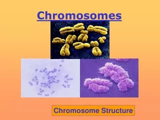
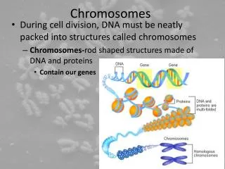
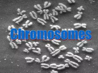
What Exactly is a chromosome? Chromosomes are the rod-shaped, filamentous bodies present in the nucleus, which become visible during cell division. They are the carriers of the gene or unit of heredity. Chromosome are not visible in active nucleus due to their high water content, but are clearly seen during cell division.
Chromosomes are composed of thin chromatin threads called Chromatin fibers. These fibers undergo folding, coiling and supercoilingduring prophase so that the chromosomes become progressively thicker and smaller. Therefore, chromosomes become readily observable under light microscope. At the end of cell division, on the other hand, the fibers uncoil and extend as fine chromatin threads, which are not visible at light microscope • Their number can be counted easily only during mitotic metaphase.
Classification of Chromosomes according to • In order to understand chromosomes and their function, we need to be able to discriminate among different chromosomes. • Size of the chromosomes • Number of chromosomes • Morphological of the chromosomes
First, Chromosome Size • In contrast to other cell organelles, the size of chromosomes shows a remarkable variation depending upon the stages of cell division. • Interphase: chromosome are longest & thinnest • Prophase: there is a progressive decrease in their length accompanied with an increase in thickness • Metaphase: Chromosomes are the most easily observed and studied during metaphase when they are very thick, quite short and well spread in the cell. • Anaphase: chromosomes are smallest. Therefore, chromosomes measurements are generally taken during mitotic metaphase.
chromosomes differ greatly in size between organisms, the size difference can be over 100-fold, while within a sp, some chromosomes are often 10 times as large as others. The size of the chromosomes in mitotic phase of animal and plants sp generally varies between 0.5 µ and 32 µ in length, and between 0.2 µ and 3.0 µ in diameter. The longest metaphase chromosomes found in Trillium- 32 µ. The giant chromosomes found in dipteraand they may be as long as 300 µ and up to 10 µ in diameter. In general, plants have longer chromosomes than animal and species having lower chromosome numbers have long chromosomes than those having higher chromosome numbers Among plants, dicotsin general, have a higher number of chromosome than monocots.Chromosomes are longer in monocot than dicots.
Karyotype&Idiotype • In a species Karyotype, a pictorial or photographic representation of all the different chromosomes in a cell of an individual, chromosomes are usually ordered by size and numbered from largest to smallest • Karyotype: is the general morphology of the somatic chromosome. Generally, karyotypes represent by arranging in the descending order of size keeping their centromeres in a straight line. • Idiotype: the karyotype of a species may be represented diagrammatically, showing all the morphological features of the chromosome; such a diagram is known as Idiotype.
Second, Number of chromosomes • Normally, all the individuals of a species have the same number of chromosomes. • Closely related species usually have similar chromosome numbers. • Presence of a whole sets of chromosomes is called euploidy. • It includes haploids, diploids, triploids, tetraploids etc. • Gametes normally contain only one set of chromosome – this number is called Haploid • Somatic cells usually contain two sets of chromosome - 2n : Diploid
Organism No. chromosomes • Human 46 • Chimpanzee 48 • Dog 78 • Horse 64 • Chicken 78 • Goldfish 94 • Fruit fly 8 • Mosquito 6 • Nematode 11(m), 12(f) • Horsetail 216 • Sequoia 22 • Round worm 2
Organism No. chromosomes • Onion 16 • Mold 16 • Carrot 20 • Tomato 24 • Tobacco 48 • Rice 24 • Maize 20 • Haploppus gracilis 4 • Crepis capillaris 6
On the extreme, round worm shows only two chromosomes, while the other extreme is represented by Protozoa having 300 or more chromosomes. • However, most organisms have numbers between 12 to 50. • 3-8 in fungi • From 8 – 16 in Angiosperms (Most common number being 12).
CHROMOSOMAL STRUCTURE A chromatid is a condensed DNA subunit of a chromosome. The two chromatids of a duplicated chromosome are held together at a region of DNA called the centromere (see figure below). Centromeres are the attachment points for microtubules, which are responsible for the guiding the movement of chromosomes during mitosis and meiosis
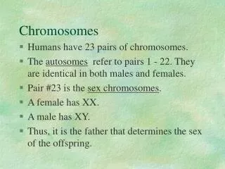
CHROMOSOMAL STRUCTURE The nuclei of most human cells contain 46 chromosomes. These 46 chromosomes consist of 23 pairs of homologous chromosomes, or homologs, meaning each of these pairs are alike, but not necessarily identical. The convention is to describe the chromosome number in humans as 2n = 46 because the cells are diploid, meaning they have two complete sets of chromosomes.
Chromosomes may differ in the position of the Centromere, the place on the chromosome where spindle fibers are attached during cell division. • The centromere divides the chromosome into two arms, • In general, if the centromere is near the middle, the chromosome is metacentric If the centromere is toward one end, the chromosome is acrocentricor submetacentric If the centromere is very near the end, the chromosome is telocentric Acrocentric Telocentric Submetacentric Metacentric
According to the position of the centromere, the eukaryotic chromosomes may be - rod-shaped (telocentric and acrocentric), - J-shaped (submetacentric) and - V-shaped (metacentric) All house mouse chromosomes are telocentric, while human chromosomes include both metacentric and acrocentric, but no telocentric.
Autosomal pair Sex chromosome DiploidNo. of No. of X Y (2n) metacentrics acrocentric or telocentric Cat 38 16 2 M M Dog 78 0 38 M A Pig 38 12 6 M M Goat 60 0 29 A M Sheep 54 3 23 A M Cow 60 0 29 M M Horse 64 13 18 M A M – Metacentric; A – Acrocentric
Third , Morphology of Eukaryotic Chromosomes Beside centromere, the chromosomes may chromosomes contain an additional specialized segment, the nucleolus organizer, which is associated with the nucleolus. A. Nucleolus organizer B. Chromosome C. Nucleolus
Satellite DNAs When the DNA of a prokaryote, such as E.coli, is isolated, fragmented and centrifuged to equilibrium in a Cesium chloride (CsCl) density gradient, the DNA usually forms a single band in the gradient. On the other hand, CsCl density-gradient analysis of DNA from eukaryotes usually reveals the presence of one large band of DNA (usually called “Mainband” DNA) and one to several small bands. These small bands are referred to as “Satellite DNAs”. For e.g., in Drosophila virilis, contain three distinct satellite DNAs. 1.Secondary constrriction2.Satellite3.Primary constriction or centromere
Morphology of Eukaryotic Chromosomes 1.Two chromonemata2.Satellite3.Centromere According to the classical cytological studies, each chromosome structurally consists of two very -thin, highly coiled filaments called chromonema or chromonemata. Each chromonemata is 800A0 thick and contains 8-microfibriis, each of which in its turn contains two double helices of DNA. Both chromonematae remain coiled in spiral manner with each other and have a series of microscopically visible bead-like swelling along its length called chromomeres.
The early geneticists have attached great significance to the chromomeres and considered them as hereditary unit, hereditary or Mendelian factors or genes; but modern cytological investigations have confirmed that the chromomeres are not genes but the regions of super-imposed coils.
Centromeres and Telomeres • Centromeres and telomeres are two essential features of all eukaryotic chromosomes. • Each provide a unique function i.e., absolutely necessary for the stability of the chromosome. • Centromeres are required for the segregation of the centromere during meiosis and mitosis. • Teleomeresprovide terminal stability to the chromosome and ensure its survival
Centromere • The region where two sister chromatids of a chromosome appear to be joined or “held together” during mitatic metaphase is called Centromere • When chromosomes are stained they typically show a dark-stained region that is the centromere. • Also termed as Primary constriction
Centromere • During mitosis, the centromere that is shared by the sister chromatids must divide so that the chromatids can migrate to opposite poles of the cell. • On the other hand, during the first meiotic division the centromere of sister chromatids must remain intact whereas during meiosis II they must act as they do during mitosis. • Therefore the centromere is an important component of chromosome structure and segregation. • As a result, centromeres are the first parts of chromosomes to be seen moving towards the opposite poles during anaphase. • The remaining regions of chromosomes lag behind and appear as if they were being pulled by the centromere.
Kinetochore • Within the centromere region, most species have several locations where spindle fibers attach, and these sites consist of DNA as well as protein. • The actual location where the attachment occurs is called the kinetochoreand is composed of both DNA and protein. • The DNA sequence within these regions is called CEN DNA.
Typically CEN DNA is about 120 base pairs long and consists of several sub-domains, CDE-I, CDE-II and CDE-III. • Mutations in the first two sub-domains have no effect upon segregation, • but a point mutation in the CDE-III sub-domain completely eliminates the ability of the centromere to function during chromosome segregation. • Therefore CDE-III must be actively involved in the binding of the spindle fibers to the centromere.
Protein complex of the kinetochore • The protein component of the kinetochore is only now being characterized. • A complex of three proteins called Cbf-III binds to normal CDE-III regions but can not bind to a CDE-III region with a point mutation that prevents mitotic segregation.
Telomere • The two ends of a chromosome are known as telomeres. • It required for the replication and stability of the chromosome. • When telomeres are damaged or removed due to chromosome breakage, the damaged chromosome ends can readily fuse or unite with broken ends of other chromosome. • Thus it is generally accepted that structural integrity and individuality of chromosomes is maintained due to telomeres.
McClintock noticed that chromosomes have unique features. • McClintock noticed that if two chromosomes were broken in a cell, the end of one could attach to the other and vice versa. • What she never observed was the attachment of the broken end to the end of an unbroken chromosome. • Thus the ends of broken chromosomes are sticky, whereas the normal end is not sticky, suggesting the ends of chromosomes have unique features.
Telomere Repeat Sequences Until recently, little was known about molecular structure of telomeres. However, during the last few years, telomeres have been isolated and characterized from several sp.
Tetrahymena- protozoa organism. • The telomeres of this organism end in the sequence 5'-TTGGGG-3'. • The telomerase adds a series of 5'-TTGGGG-3' repeats to the ends of the lagging strand. • A hairpin occurs when unusual base pairs between guanine residues in the repeat form. • Finally, the hairpin is removed at the 5'-TTGGGG-3' repeat. • Thus the end of the chromosome is faithfully replicated. RNA Primer - Short stretches of ribonucleotides (RNA substrates) found on the lagging strand during DNA replication. Helps initiate lagging strand replication