Raman Spectroscopy Chapter 18
Raman Spectroscopy Chapter 18 . Dr. Nizam M. El-Ashgar Chemistry Department Islamic University of Gaza. Ch 18 Raman Spectroscopy. When radiation passes through a transparent medium, the species present scatter a fraction of the beam in all directions.

Raman Spectroscopy Chapter 18
E N D
Presentation Transcript
Raman SpectroscopyChapter 18 Dr. Nizam M. El-Ashgar Chemistry Department Islamic University of Gaza
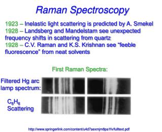
Ch 18Raman Spectroscopy • When radiation passes through a transparent medium, the species present scatter a fraction of the beam in all directions. • In 1928, the Indian physicist C. V. Raman discovered that: • The visible wavelength of a small fraction of the radiation scattered by certain molecules differs from that of the incident beam. • Furthermore that the shifts in wavelength depend upon the chemical structure of the molecules responsible for the scattering.
November 7, 1888-November 21, 1970 Won the Noble Prize in 1930 for Physics Discovered the "Raman Effect" Besides Discovering the Raman Effect, He studied extensively in X-Ray Diffractions, Acoustics, Optics, Dielectrics, Ultrasonics, Photo electricity, and colloidal particles. Sir Chandrashekhara Venkata Raman
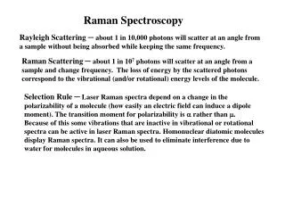
Raman Spectroscopy • The theory of Raman scattering shows that the phenomenon results from the same type of quantized vibrational changes that are associated with infrared absorption. • Thus, the difference in wavelength between the incident and scattered visible radiation corresponds to wavelengths in the mid-infrared region. • The Raman scattering spectrum and infrared absorption spectrum for a given species often resemble one another quite closely.
Raman Spectroscopy • An important advantage of Raman spectra over infrared lies in the fact that water does not cause interference; indeed, Raman spectra can be obtained from aqueous solutions. • In addition, glass or quartz cells can be employed, thus avoiding the inconvenience of working with sodium chloride or other atmospherically unstable window materials.
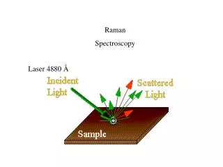
THEORY OF RAMAN SPECTROSCOPY • Raman spectra are acquired by irradiating a sample with a powerful laser source of visible or near-infrared monochromatic radiation. • During irradiation, the spectrum of the scattered radiation is measured at some angle (often 90 deg) with a suitable spectrometer. • At the very most, the intensities of Raman lines are 0.001 % of the intensity of the source; as a consequence, their detection and measurement are somewhat more difficult than are infrared spectra.
Light Scattering Phenomenon • When radiation passes through a transparent medium, the species present in that medium scatter a fraction of the beam in all directions.
Scattering of Radiation • The fraction of radiation transmitted at all angles from its original path. • Expalanation: • Transmission of radiation in matter can be pictured as: • A momentary retention of the radiant energy by atoms, ions, or molecules followed by reemission of the radiation in all directions as the particles return to their original state. • With atomic or molecular particles that are small relative to the wavelength of the radiation, destructive interference removes most but not all of the reemitted radiation except the radiation that travels in the original direction of the beam; the path of the beam appears to be unaltered as a consequence of the interaction.
Careful observation, however, reveals that: A very small fraction of the radiation is transmitted at all angles from the original path and that the intensity of this scattered radiation increases with particle size. Types of Scattering: • Rayleigh scattering Scattering by molecules or aggregates of molecules with dimensions significantly smaller than the wavelength of the radiation. Its intensity is proportional to: • The inverse fourth power of the wavelength (. • The dimensions of the scattering particles. • The square of the polarizability of the particles. • An everyday manifestation of Rayleigh scattering is the blue color of the sky, which results from greater scattering of the shorter wavelengths of the visible spectrum.
The blue color of the sky is caused by the scattering of sunlight off the molecules of the atmosphere. This scattering, called, is more effective at short wavelengths (the blue end of the visible spectrum). Therefore the light scattered down to the earth at a large angle with respect to the direction of the sun's light is predominantly in the blue end of the spectrum.
Scattering by Large Molecules : • With particles of colloidal diamensions, scattering is sufficiently intense to be seen by naked eye (Tyndal effect). • Used to determine the size and shape of polymer molecules and colloidal particles. Raman Scattering • The Raman scattering effect differs from ordinary scattering in that part of the scattered radiation suffers quantized frequency changes. • These changes are the result of vibrational energy level transitions that occur in the molecules as a consequence of the polarization process.
Basic Physical Realization • Illuminate a specimen with laser light (e.g. 532nm) • Scattered (no absorbed) Light in two forms • Elastic (Rayleigh) → scattered = incident • InElastic (Raman) → scattered incident • Light Experiences a “Raman Shift” in Wave Length.
Raman vs. Rayleigh • Raman Intensity is About 0.1 ppm of the Incoming Laser Intensity
Raman Stokes Scattering Raman AntiStokes Scattering • Inelastic Light Scattering Mechanisms • Raman Shift Can be: • To Longer WaveLengths (Stokes Scattering) • Loses Energy – Predominant Raman Shift • To Shorter WaveLengths (AntiStokes Scattering) • Gains Energy – Subordinate Raman Shift
Raman Spectroscopy: photon interpretation • Raman effect is a 2-photon scattering process • These processes are inelastic scattering: • Stokes scattering: energy lost by photon: • — (( — )) Photon in Photon out No vibration Vibration • Anti-Stokes scattering: energy gained by photon: (( — )) — Photon in Photon out Vibration No vibration
But dominant process is elastic scattering: • Rayleigh scattering • — — Photon in Photon out No vibration No vibration If incident photon energy E; vibration energy v, then in terms of energy, photon out has energy: E-v Stokes scattering E+v anti-Stokes scattering E Rayleigh scattering
Representation in terms of energy levels: Arrow up = laser photon in; Arrow down = Raman scattering out
Excitation of Raman Spectra: Summery A Raman spectrum can be obtained by irradiating a sample of carbon tetrachloride (Fig 18-2) with an intense beam of an argon ion laser having a wavelength of 488.0 nm (20492 cm-1). The emitted radiation is of three types: 1. Stokes scattering 2. Anti-stokes scattering 3. Rayleigh scattering
Excitation of Raman Spectra • The abscissa of Raman spectrum is the wavenumber shift , which is defined as the difference in wavenumbers (cm-1) between the observed radiation and that of the source. • For CCl4 three peaks are found on both sides of the Rayleigh peak and that the pattern of shifts on each side is identical (Fig. 18-2). • Anti-Stokes lines are appreciably less intense than the corresponding Stokes lines. For this reason, only the Stokes part of a spectrum is generally used. • The magnitude of Raman shifts are independent of the wavelength of excitation.
Mechanism of Raman and Rayleigh Scattering • The heavy arrow on the far left (bold black “up” arrow) depicts the energy change in the molecule when it interacts with a photon. • The increase in energy is equal to the energy of the photon h. • The second and narrower arrow (thin black “up” arrow) shows the type of change that would occur if the molecule is in the first vibrational level of the electronic ground state.
Mechanism of Raman and Rayleigh Scattering • The middle set of arrows (the first two blue arrows) depicts the changes that produce Rayleigh scattering. • The energy changes that produce stokes and anti-Stokes emission are depicted on the right (the last two blue arrows). • The two differ from the Rayleigh radiation by frequencies corresponding to E, the energy of the first vibrational level of the ground state. • If the bond were infrared active, the energy of its absorption would also be E. • Thus, the Raman frequency shift and the infrared absorption peak frequency are identical.
Mechanism of Raman and Rayleigh Scattering • The relative populations of the two upper energy states are such that Stokes emission is much favored over anti-Stokes. • Rayleigh scattering has a considerably higher probability of occurring than Raman because the most probable event is the energy transfer to molecules in the ground state and reemission by the return of these molecules to the ground state. • The ratio of anti-Stokes to Stokes intensities will increase with temperature because a larger fraction of the molecules will be in the first vibrationally excited state under these circumstances.
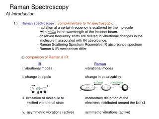
Raman vs. I.R • For a given bond, the energy shifts observed in a Raman experiment should be identical to the energies of its infrared absorption bands, • provided that the vibrational modes involved are active toward both infrared absorption and Raman scattering. • The differences between a Raman spectrum and an infrared spectrum are not surprising. • Infrared absorption requires that a vibrational mode of the molecule have a change in dipole moment or charge distribution associated with it.
Raman vs. I.R • In contrast, scattering involves a momentary distortion of the electrons distributed around a bond in a molecule, followed by reemission of the radiation as the bond returns to its normal state. • In its distorted form, the molecule is temporarily polarized; that is, it develops momentarily an induced dipole that disappears upon relaxation and reemission. • The Raman activity of a given vibrational mode may differ markedly from its infrared activity.
Selection rule for Raman spectrum Vibration is active if it has a change in polarizability, . Polarizability is the ease of distortion of a bond. For Raman-active vibrations, the incident radiation does not cause a change in the dipole moment of the molecule, but instead a change in polarizability. In starting the vibration going, the electric field of the radiation at time t, E, induces a separation of charge (i.e. between the nuclear protons and the bonding electrons). This is called the induced dipole moment, P. (Don’t confuse it with the molecule’s dipole moment, or change in dipole moment, because this is often zero). P = E
Factors affect Polarizability 1- Atomic number Z: P the amount of electrons, Electrons become less control by nuclear charge. 2- Bond Length: P Bond Length 3- Atomic or Molecular Size: P Size, 4- Molecular orientation with respect to an electric field Parallel or perpendicular (Exp: Parallel has more effect) 5- Bond Strength (Bond order): P 1/strength of bondC=C, and C≡C, C≡N bonds are strong scatterers, bonds undergo polarization. 6- Electronegativity difference: P 1/ difference in electronegativity 7- Covalent bonds more polarizable than ionic bonds.
Raman Scattering is Stronger from Some Vibrations than from Others • Stretching bands often stronger than bending ones • Symmetric bands often stronger than anti-symmetric ones: • Symmetric stretches undergo greater changes in polarization, and are stronger in Raman than asymmetric stretches. • Crystalline materials often have stronger Raman bands than non-crystalline materials
Homoneuclear molecules such as Cl2 , N2 and , H2 , are polarizable(vary periodically in phase with the stretching vibrations and increases with separation), so they are Raman active. • For molecules with a center of symmetry, no IR active transitions are Raman active and vice versa • Symmetric molecules IR-active vibrations are not Raman-active. Raman-active vibrations are not IR-active. O = C = O O = C = O Raman active Raman inactive IR inactive IR active
Water Symmetrical stretch Asymmetric stretch Bending mode IR active: change of dipole moment & Raman active: change in electronic polarizability
O=C=O O=C=O Raman-active and Non-Raman-active Vibrations (II) Symmetric Stretching of CO2 1=1340 cm-1 No change in dipole moment – IR inactive Change in polarizability – Raman active Asymmetric Stretching of CO2 2= 2350 cm-1 Change so much in dipole moment – IR active Non-change in polarizability – Raman inactive
Raman-active and Non-Raman-active Vibrations (III) Symmetric Stretching of CS2 1 S=C=S No change in dipole moment – non-IR activity Change in polarizability – Raman activity
S=C=S Raman-active and Non-Raman-active Vibrations (IV) Asymmetric Stretching of CS2 2 Change so much in dipole moment – IR activity Non-change in polarizability – Raman inactivity
S=C=S Raman-active and Non-Raman-active Vibrations (V) Bending of CS2 3 Change so much in dipole moment – IR activity Non-change in polarizability – Raman inactivity
Intensity of Normal Raman Peaks • The intensity or power of a normal Raman peak depends in a complex way upon • The polarizability of the molecule: • The intensity of the source, • The concentration of the active group. • The power of Raman emission increases with the fourth power of the frequency of the source; however, advantage can seldom be taken of this relationship because of the likelihood that ultraviolet irradiation will cause photodecomposition. • Raman intensities are usually directly proportional to the concentration of the active species.
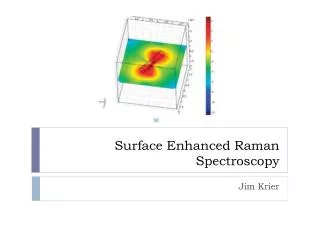
Raman Depolarization Ratios Polarizability: describes a molecular property having to do with the deformability of a bond. • Polarization: is a property of a beam of radiation and describes the plane in which the radiation vibrates. • Raman spectra are excited by plane-polarized radiation. • The scattered radiation is found to be polarized to various degrees depending upon the type of vibration responsible for the scattering.
z y x Light Scattering, Depolarization Ratio & Molecular Orientation (I) Asymmetric Sample Incident laser Polarimeter
When Raman spectra are excited by plane-polarized radiation, by using a laser source, the scattered radiation is found to be polarized to various degrees depending on the type of vibration responsible for the scattering. The nature of this effect is illustrated in the previous Figure, where radiation from a laser source is shown as being polarized in the yz plane. Part of the resulting scattered radiation is shown as being polarized parallel to the original beam, that is, in the xz plane; the intensity of this radiation is symbolized by the subscript ║. The remainder of the scattered beam is polarized in the xy plane, which is perpendicular to the polarization of the original beam; the intensity of this perpendicularly polarized radiation is shown by the subscript ┴.
Raman Depolarization Ratios The depolarization ratio p is defined as Experimentally, the depolarization ratio may be obtained by inserting a polarizer between the sample and the monochromator. The depolarization ratio is dependent upon the symmetry of the vibrations responsible for scattering.
Depolarization ratio of a vibrational mode in the Raman spectrum may give information about the symmetry of a vibration. p = depolarization ratio for polarized light = Iy/Iz = I/I|| This is different from the depolarization ratio for unpolarized light, see Infrared and Raman Spectra…Part A., K. Nakamoto 5th Ed. Wiley 1997. Pp. 97-101. 0 p <0.75; Raman line is polarized (p). Vibration is totally symmetric p = 0.75. Raman line is depolarized (dp). Vibration is not totally symmetric. CCl4
Raman Depolarization Ratios Polarized band: p = < 0.75 for totally symmetric modes. Depolarized band: p = 0.75 for nonsymmetrical vibrational modes Anomalously polarized band: p = > 0.75 for vibrational modes
Raman Polarized Spectra of SO2 Asymmetric stretching vibration
Light Scattering, Depolarization Ratio & Molecular Orientation (III) 100% ρ= 75% CCl4 459cm-1 - Symmetric stretching 314, 218cm-1 - Asymmetric stretching ρ=0.007 459 314 218 Raman Shift /cm-1