Download

1 / 36
360 likes | 588 Vues
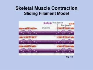
Summary of events during skeletal muscle contraction. Rest. actin and myosin uncoupled calcium stored in SR. Excitation. nerve impulse generated ACH released from the vesicles sarcolemma depolarized muscle impulse transmitted through the fiber calcium released from the cisternae

E N D
Rest • actin and myosin uncoupled • calcium stored in SR
Excitation • nerve impulse generated • ACH released from the vesicles • sarcolemma depolarized • muscle impulse transmitted through the fiber • calcium released from the cisternae • calcium binds to troponin • actin binding sites activated • myosin ATPase activated
Contraction • Myosin cross-bridges swivel • Release ADP + Pi • Actin slides over myosin
Regeneration • ATP attaches to myosin • actin and myosin dissociate • ATP → ADP + Pi • contraction process repeats
Relaxation • ACH decomposed by cholinesterase • nerve impulse stops • calcium removed by calcium pump • actin binding sites inhibited (Tn/tropomyosin complex returns to original position) • muscle returns to resting state
Excitation Contraction Coupling • process by which myofibrils translate nerve impulses into muscle contraction • depolarization of t-tubule membrane (a change in the membrane potential) results in the calcium release from the terminal cisternae of the SR • this results in muscle contraction
also know calcium is resequestered in the SR via active calcium-ATPase pumps • not clear is how the change in membrane potential of the t-tubule system is communicated into the SR to cause calcium release
Structure and Function of the Triad • t-tubules and SR communicate at the triad junction • a ryanodine receptor is a large protein complex which is the release channel of the sarcoplasmic calcium, located at the t-tubule SR junction • the receptor has two parts: channel region and a large cytoplasmic region
within t-tubule is another protein complex, believed to be the voltage sensor which controls the opening and closing of the ryanodine receptor, this is the DHP or dihydropyridine receptor complex • hypothesized: ryanodine receptor and the DHP receptor interact with each other to cause the action potential to induce calcium release from the SR
Theories of Communication Calcium-induced tubule membranes Voltage-induced changes in the t-tubule Changes in the voltage gradient
Calcium induced tubule membranes Cause an opening of the calcium channel calcium released: stimulation of t-tubule induces a release of calcium ions from the gates within the SR
Con: block calcium release channels of the cell membrane does not inhibit calcium release from the SR Nor reduce tension development Also, amount of calcium needed to induce release is not clear (may not be physiologic)
Voltage-induced changes in the t-tubule • induce the formation of D-myosinositol 1,4,5-triphosphate (IP3) • IP3 then increases the permeability of the SR membranes to cause calcium release
Pros: elevated IP3 production has been demonstrated in skeletal muscle following electrical stimulation Release of calcium in skinned fibers induced by IP3 An augmented muscle response following inhibition of IP3 breakdown A reduction of IP3 release from RBCs Cons: is the concentration of IP3 necessary for activation and is its rate of activation physiologic?
Changes in the voltage gradient • Activation of calcium is due to perturbation of the normal H+ gradient across the SR membranes • Cons: the pH changes during muscle contraction may be too small to induce the necessary gradients in order to stimulate calcium release
Hypothesized Mechanisms for ECC • Calcium-induced calcium release • Chemical intermediate • Allosteric Interaction
Calcium induced calcium release • stimulation of t-tubules causes a small amount of calcium to cross the t-tubule membrane • an amount insufficient to cause muscle contraction • This induces the release of additional calcium in greater amounts, from the SR (calcium channels are open)
Cons: blocking the DHP channels of the t-tubule membrane does not inhibit calcium release from the SR Or decrease force development Are the amounts of calcium required physiologic?
Chemical Intermediate • voltage-induced changes in the t-tubules induce the formation of inositol 1,4,5 triphosphate (InsP3) • IP3 then increases the permeability of the SR membranes to cause calcium release
Pros: after electrical stimulation, there is an elevated production IP3 The release of calcium in skinned fibers induced by IP3 If IP3 release from the RBCs is inhibited, there is a reduction of calcium transients in skeletal muscle If IP3 breakdown is inhibited, there is an augmented skeletal muscle response
Cons: is the concentration of IP3 necessary for activation? Is its rate of activation physiologic?
Allosteric Interaction Hypothesis (aka, Plunger Hypothesis) • there is a mechanical or allosteric link between DHP channels of the t-tubules and the ryanodine channels of the SR (junctional foot proteins) • There is a foot structure composed primarily of SR calcium channel proteins clustered next to the t-tubule membrane called channel protein or electron dense feet
This suggests a role for protein-protein interactions to transmit the depolarization signal across the junction. Most popular mechanism of signal transmission is a variant of the plunger hypothesis: • Imagine a mobile positive charge in the t-tubule membrane (it may be associated with the DHP voltage sensor)
The charge is connected by a rod to a plug in the calcium release channel of the SR • Depolarization of the t-tubule membrane causes charge movement in the t-tubule membrane • Results in the unplugging of the SR calcium release channel
Suspect that the mobile charge is contained within specific amino acids contained within a segment of the DHP receptor complex • Ryanodine receptor complex has a large cytoplasmic portion spanning the gap between membranes, and able to contact the voltage sensors of the DHP complex
Skeletal Muscle Fiber Types not all skeletal muscle has the same biochemical or functional characteristics are generally classified according to their primary dependence on different metabolic pathways for the production of ATP.
Nomenclature based on Myosin ATPase pH lability: histochemical staining Glycolytic staining Oxidative staining
Fiber sub-types: • Type I, SO (slow oxidative), red fibers, slow • Type IIa FOG (fast oxidative glycolytic), intermediate fibers • Type IIb FG (fast glycolytic), white, fast
Type IIc rare, undifferentiated fiber, perhaps found during re-innervation or motor unit transformation, between I and IIa on the continuum of metabolic potential • Type IIx, not classified
Slow twitch fibers: • fatigue resistant, good for prolonged exercise • primarily synthesized ATP via aerobic energy transfer • recruited for aerobic activities such as prolonged moderate exercise • have a smaller resting membrane potential, -50-70mv, versus 80-90mv in fast muscle
longer latency period due to less extensive SR • low activity of myosin ATPase • slow speed of contraction • low glycolytic capacity • increased size and number of mitochondria
higher levels of myoglobin • higher concentrations of mitochondrial enzymes • increased blood flow -- increased capillarization
Fast twitch fibers: • activated in short-term, sprint activities, and forceful contractions which rely primarily on anaerobic metabolism for energy • important in stop and go and change of pace activities • more extensive SR • greater capability for electrochemical AP transmission (due to increased SR)
high activity level of myosin ATPase • rapid SR calcium release and uptake • high rate of cross-bridge turnover development • intrinsic speed of contraction and tension is 2-3 times that of ST fibers • primarily use the glycolytic system for energy transfer