Download

1 / 22
340 likes | 905 Vues
Skeletal Muscle Contraction. Muse. Epimysium. Epimysium. Bone. Perimysium. Endomysium. Tendon. Muscle fiber in middle of a fascicle. (b). Blood vessel. Fascicle (wrapped by perimysium). Endomysium (between individual muscle fibers). Perimysium. Fascicle. Muscle fiber. (a).

E N D
Epimysium Epimysium Bone Perimysium Endomysium Tendon Muscle fiber in middle of a fascicle (b) Blood vessel Fascicle (wrapped by perimysium) Endomysium (between individual muscle fibers) Perimysium Fascicle Muscle fiber (a) Muscle cells are composed of many long fibers
Sarcolemma Mitochondrion Myofibril Dark A band Light I band Nucleus (b) Diagram of part of a muscle fiber showing the myofibrils. Onemyofibril is extended from the cut end of the fiber. Cells are polynucleate and surrounded by sarcolemma
The overlap of fiber elements causes the striated appearance Z disc Z disc
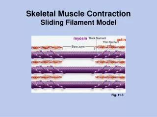
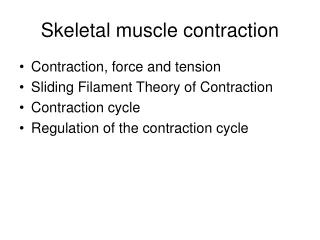
The Sarcomere • Smallest contractile unit (functional unit) of a muscle fiber • The region of a myofibril between two successive Z discs • Composed of thick and thin myofilaments made of contractile proteins
Positions of the fibers relative to each other Thin (actin) filament Z disc H zone Z disc Thick (myosin) filament I band A band Sarcomere I band M line (c) Small part of one myofibril enlarged to show the myofilaments responsible for the banding pattern. Each sarcomereextends from one Z disc to the next. Sarcomere Z disc Z disc M line Thin (actin) filament Elastic (titin) filaments Thick (myosin) filament (d) Enlargement of one sarcomere (sectioned lengthwise). Notice the myosin heads on the thick filaments. Thin filaments = actin Thick filaments = myosin
Myosin is a tree of heads Myosin thick filament
The signal to contract 10_10 Action potential arrives at neuromuscular junction. acetocholine released ligand gated sodium channels open and action potential continues along sarcolemma
1 Action potential is propagated along the sarcolemma and down the T tubules. Steps in E-C Coupling: Sarcolemma Voltage-sensitive tubule protein T tubule Ca2+ release channel Terminal cisterna of SR Ca2+ Figure 9.11, step 3
Part of a skeletal muscle fiber (cell) I band A band I band Z disc H zone Z disc Myofibril M line Sarcolemma Triad: T tubule • • Terminal cisternae of the SR (2) Sarcolemma Tubules of the SR Myofibrils Mitochondria Sarcoplasmic reticulum releases Ca2+
The Contraction Cycle • Five Steps of the Contraction Cycle • Exposure of active sites on actin • Formation of cross-bridges • Pivoting of myosin heads • Detachment of cross-bridges • Reactivation of myosin
The length of the sarcomere shrinks as the filaments undergo the contraction cycle
Once detached, residual energy from detachment re-cocks myosin head
review 10_11
Different fiber types can be fast or slower to develop full tension .