Download

1 / 52
530 likes | 628 Vues
Explore the intricate details of the kidney's external and internal anatomy, its crucial role in regulating blood composition and pH, and the complex nephron structures. Understand the renal physiology processes like glomerular filtration, tubular reabsorption, and secretion. Discover the significance of nephrons, renal corpuscles, and collecting ducts in maintaining kidney health.

E N D
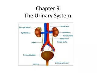
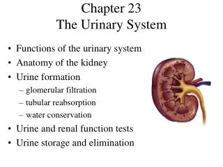
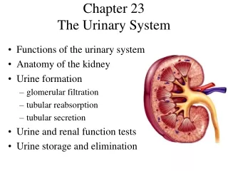
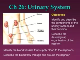
Chapter 26The Urinary System • Kidneys, ureters, urinary bladder & urethra • Urine flows from each kidney, down its ureter to the bladder and to the outside via the urethra • Filter the blood and return most of water and solutes to the bloodstream
Overview of Kidney Functions • Regulation of blood ionic composition • Na+, K+, Ca+2, Cl- and phosphate ions • Regulation of blood pH, osmolarity & glucose • Regulation of blood volume • conserving or eliminating water • Regulation of blood pressure • secreting the enzyme renin • adjusting renal resistance • Release of erythropoietin & calcitriol • Excretion of wastes & foreign substances
External Anatomy of Kidney • Paired kidney-bean-shaped organ • 4-5 in long, 2-3 in wide,1 in thick • Found just above the waist between the peritoneum & posterior wall of abdomen • retroperitoneal along with adrenal glands & ureters • Protected by 11th & 12th ribs with right kidney lower
External Anatomy of Kidney • Blood vessels & ureter enter hilus of kidney • Renal capsule = transparent membrane maintains organ shape • Adipose capsule that helps protect from trauma • Renal fascia = dense, irregular connective tissue that holds against back body wall
Internal Anatomy of the Kidneys • Parenchyma of kidney • renal cortex = superficial layer of kidney • renal medulla • inner portion consisting of 8-18 cone-shaped renal pyramids separated by renal columns • renal papilla point toward center of kidney • Drainage system fills renal sinus cavity • cuplike structure (minor calyces) collect urine from the papillary ducts of the papilla • minor & major calyces empty into the renal pelvis which empties into the ureter
Internal Anatomy of Kidney • What is the difference between renal hilus & renal sinus? • Outline a major calyx & the border between cortex & medulla.
Blood & Nerve Supply of Kidney • Abundantly supplied with blood vessels • receive 25% of resting cardiac output via renal arteries • Functions of different capillary beds • glomerular capillaries where filtration of blood occurs • vasoconstriction & vasodilation of afferent & efferent arterioles produce large changes in renal filtration • peritubular capillaries that carry away reabsorbed substances from filtrate • Sympathetic vasomotor nerves regulate blood flow & renal resistance by altering arterioles
Blood Vessels around the Nephron • Glomerular capillaries are formed between the afferent & efferent arterioles • Efferent arterioles give rise to the peritubular capillaries and vasa recta
The Nephron • Kidney has over 1 million nephrons composed of a corpuscle and tubule • Renal corpuscle = site of plasma filtration • glomerulus is capillaries where filtration occurs • glomerular (Bowman’s) capsule is double-walled epithelial cup that collects filtrate • Renal tubule • proximal convoluted tubule • loop of Henle dips down into medulla • distal convoluted tubule • Collecting ducts and papillary ducts drain urine to the renal pelvis and ureter
Cortical Nephron • 80-85% of nephrons are cortical nephrons • Renal corpuscles are in outer cortex and loops of Henle lie mainly in cortex
Juxtamedullary Nephron • 15-20% of nephrons are juxtamedullary nephrons • Renal corpuscles close to medulla and long loops of Henle extend into deepest medulla enabling excretion of dilute or concentrated urine
Histology of the Nephron & Collecting Duct • Single layer of epithelial cells forms walls of entire tube • Distinctive features due to function of each region • microvilli • cuboidal versus simple • hormone receptors
Structure of Renal Corpuscle • Bowman’s capsule surrounds capsular space • podocytes cover capillaries to form visceral layer • simple squamous cells form parietal layer of capsule • Glomerular capillaries arise from afferent arteriole & form a ball before emptying into efferent arteriole
Juxtaglomerular Apparatus • Structure where afferent arteriole makes contact with ascending limb of loop of Henle • macula densa is thickened part of ascending limb • juxtaglomerular cells are modified muscle cells in arteriole
Number of Nephrons • Remains constant from birth • any increase in size of kidney is size increase of individual nephrons • If injured, no replacement occurs • Dysfunction is not evident until function declines by 25% of normal (other nephrons handle the extra work) • Removal of one kidney causes enlargement of the remaining until it can filter at 80% of normal rate of 2 kidneys
Overview of Renal Physiology • Nephrons and collecting ducts perform 3 basic processes • glomerular filtration • a portion of the blood plasma is filtered into the kidney • tubular reabsorption • water & useful substances are reabsorbed into the blood • tubular secretion • wastes are removed from the blood & secreted into urine • Rate of excretion of any substance is its rate of filtration, plus its rate of secretion, minus its rate of reabsorption
Overview of Renal Physiology • Glomerular filtration of plasma • Tubular reabsorption • Tubular secretion
Glomerular Filtration • Blood pressure produces glomerular filtrate • Filtration fraction is 20% of plasma • 48 Gallons/dayfiltrate reabsorbedto 1-2 qt. urine • Filtering capacityenhanced by: • thinness of membrane & large surface area of glomerular capillaries • glomerular capillary BP is high due to small size of efferent arteriole
Filtration Membrane • #1 Stops all cells and platelets • #2 Stops large plasma proteins • #3 Stops medium-sized proteins, not small ones
Glomerular Filtration Rate • Amount of filtrate formed in all renal corpuscles of both kidneys / minute • average adult male rate is 125 mL/min • Homeostasis requires GFR that is constant • too high & useful substances are lost due to the speed of fluid passage through nephron • too low and sufficient waste products may not be removed from the body • Changes in net filtration pressure affects GFR • filtration stops if GBHP drops to 45mm Hg • functions normally with mean arterial pressures 80-180
Renal Autoregulation of GFR • Mechanisms that maintain a constant GFR despite changes in arterial BP • myogenic mechanism • systemic increases in BP, stretch the afferent arteriole • smooth muscle contraction reduces the diameter of the arteriole returning the GFR to its previous level in seconds • tubuloglomerular feedback • elevated systemic BP raises the GFR so that fluid flows too rapidly through the renal tubule & Na+, Cl- and water are not reabsorbed • macula densa detects that difference & releases a vasoconstrictor from the juxtaglomerular apparatus • afferent arterioles constrict & reduce GFR
Neural Regulation of GFR • Blood vessels of the kidney are supplied by sympathetic fibers that cause vasoconstriction of afferent arterioles • At rest, renal BV are maximally dilated because sympathetic activity is minimal • renal autoregulation prevails • With moderate sympathetic stimulation, both afferent & efferent arterioles constrict equally • decreasing GFR equally • With extreme sympathetic stimulation (exercise or hemorrhage), vasoconstriction of afferent arterioles reduces GFR • lowers urine output & permits blood flow to other tissues
Tubular Reabsorption & Secretion • Normal GFR is so high that volume of filtrate in capsular space in half an hour is greater than the total plasma volume • Nephron must reabsorb 99% of the filtrate • PCT with their microvilli do most of work with rest of nephron doing just the fine-tuning • solutes reabsorbed by active & passive processes • water follows by osmosis • small proteins by pinocytosis • Important function of nephron is tubular secretion • transfer of materials from blood into tubular fluid • helps control blood pH because of secretion of H+ • helps eliminate certain substances (NH4+, creatinine, K+)
Transport Mechanisms • Water is only reabsorbed by osmosis • obligatory water reabsorption occurs when water is “obliged” to follow the solutes being reabsorbed • facultative water reabsorption occurs in collecting duct under the control of antidiuretic hormone
Glucosuria • Common cause is diabetes mellitis because insulin activity is deficient and blood sugar is too high
Reabsorption in the Loop of Henle • Tubular fluid • PCT reabsorbed 65% of the filtered water so chemical composition of tubular fluid in the loop of Henle is quite different from plasma • since many nutrients were reabsorbed as well, osmolarity of tubular fluid is close to that of blood • Sets the stage for independent regulation of both volume & osmolarity of body fluids
Symporters in the Loop of Henle • Thick limb of loop of Henle has Na+ K- Cl- symporters that reabsorb these ions • K+ leaks through K+ channels back into the tubular fluid leaving the interstitial fluid and blood with a negative charge • Cations passively move to the vasa recta
Reabsorption & Secretion in the Collecting Duct • By end of DCT, 95% of solutes & water have been reabsorbed and returned to the bloodstream • Cells in the collecting duct make the final adjustments • principal cells reabsorb Na+ and secrete K+ • intercalated cells reabsorb K+ & bicarbonate ions and secrete H+
Actions of the Principal Cells • Na+ enters principal cellsthrough leakage channels • Na+ pumps keep theconcentration of Na+ inthe cytosol low • Cells secrete variableamounts of K+, to adjustfor dietary changes in K+intake • down concentration gradient due to Na+/K+ pump • Aldosterone increases Na+ and water reabsorption & K+ secretion by principal cells by stimulating the synthesis of new pumps and channels.
Secretion of H+ and Absorption of Bicarbonate by Intercalated Cells • Proton pumps (H+ATPases) secrete H+ into tubular fluid • can secrete against a concentration gradient so urine can be 1000 times more acidic than blood
Hormonal Regulation • Hormones that affect Na+, Cl- & water reabsorption and K+ secretion in the tubules • angiotensin II and aldosterone • decreases GFR by vasoconstricting afferent arteriole • enhances absorption of Na+ • promotes aldosterone production which causes principal cells to reabsorb more Na+ and Cl- and less water • increases blood volume by increasing water reabsorption
Antidiuretic Hormone • Increases water permeability of principal cells • When osmolarity of plasma & interstitial fluid decreases, more ADH is secreted
Production of Dilute or Concentrated Urine • Homeostasis of body fluids despite variable fluid intake • Kidneys regulate water loss in urine • ADH controls whether dilute or concentrated urine is formed • if lacking, urine contains high ratio of water to solutes
Formation of Dilute Urine • Dilute = having fewer solutes than plasma • diabetes insipidus • Filtrate and blood have equal osmolarity in PCT • Principal cells do not reabsorb water if ADH is low
Formation of Concentrated Urine • Compensation for low water intake or heavy perspiration • Urine can be up to 4 times greater osmolarity than plasma • Cells in the collecting ducts reabsorb more water & urea when ADH is increased
Summary • H2O Reabsorption • PCT---65% • loop---15% • DCT----10-15% • collecting duct--- 5-10% with ADH • Dilute urine has not had enough water removed, although sufficient ions have been reabsorbed.
Diuretics • Substances that slow renal reabsorption of water & cause diuresis (increased urine flow rate) • caffeine which inhibits Na+ reabsorption • alcohol which inhibits secretion of ADH • prescription medicines can act on the PCT, loop of Henle or DCT
Evaluation of Kidney Function • Urinalysis • analysis of the volume and properties of urine • normal urine is protein free, but includes filtered & secreted electrolytes • urea, creatinine, uric acid, urobilinogen, fatty acids, enzymes & hormones • Blood tests • blood urea nitrogen test (BUN) measures urea in blood • rises steeply if GFR decreases severely • plasma creatinine--from skeletal muscle breakdown • renal plasma clearance of substance from the blood in ml/minute (important in drug dosages)
Dialysis Therapy • Kidney function is so impaired the blood must be cleansed artificially • separation of large solutes from smaller ones by a selectively permeable membrane • Artificial kidney machine performs hemodialysis • directly filters blood because blood flows through tubing surrounded by dialysis solution • cleansed blood flows back into the body
Anatomy of Ureters • 10 to 12 in long • Varies in diameter from 1-10 mm • Extends from renal pelvis to bladder • Retroperitoneal • Enters posterior wall of bladder • Physiological valve only • bladder wall compresses arterial opening as it expands during filling • flow results from peristalsis, gravity & hydrostatic pressure
Histology of Ureters • 3 layers in wall • mucosa is transitional epithelium & lamina propria • since organ must inflate & deflate • mucus prevents the cells from being contacted by urine • muscularis • inner longitudinal & outer circular smooth muscle layer • distal 1/3 has additional longitudinal layer • peristalsis contributes to urine flow
Location of Urinary Bladder • Posterior to pubic symphysis • In females is anterior to vagina & inferior to uterus • In males lies anterior to rectum
Anatomy of Urinary Bladder • Hollow, distensible muscular organ with capacity of 700 - 800 mL • Trigone is smooth flat area bordered by 2 ureteral openings and one urethral opening
Histology of Urinary Bladder • 3 layers in wall • mucosa is transitional epithelium & lamina propria • since organ must inflate & deflate • mucus prevents the cells from being contacted by urine • muscularis (known as detrusor muscle) • 3 layers of smooth muscle • inner longitudinal, middle circular & outer longitudinal • circular smooth muscle fibers form internal urethral sphincter • circular skeletal muscle forms external urethral sphincter • adventitia layer of loose connective tissue anchors in place • superior surface has serosal layer (visceral peritoneum)
Micturition Reflex • Micturition or urination (voiding) • Stretch receptors signal spinal cord and brain • when volume exceeds 200-400 mL • Impulses sent to micturition center in sacral spinal cord (S2 and S3) & reflex is triggered • parasympathetic fibers cause detrusor muscle to contract, external & internal sphincter muscles to relax • Filling causes a sensation of fullness that initiates a desire to urinate before the reflex actually occurs • conscious control of external sphincter • cerebral cortex can initiate micturition or delay its occurrence for a limited period of time
Anatomy of the Urethra • Females • length of 1.5 in., orifice between clitoris & vagina • histology • transitional changing to nonkeratinized stratified squamous epithelium, lamina propria with elastic fibers & circular smooth muscle • Males • tube passes through prostate, UG diaphragm & penis • 3 regions of urethra • prostatic urethra, membranous urethra & spongy urethra • circular smooth muscle forms internal urethral sphincter & UG diaphragm forms external urethral sphincter
Urinary Incontinence • Lack of voluntary control over micturition • normal in 2 or 3 year olds because neurons to sphincter muscle is not developed • Stress incontinence in adults • caused by increases in abdominal pressure that result in leaking of urine from the bladder • coughing, sneezing, laughing, exercising, walking • injury to the nerves, loss of bladder flexibility, or damage to the sphincter