Download
1 / 17
170 likes | 233 Vues
Learn about the components and functions of the urinary system, nephron organization, blood flow, kidney anatomy, urine collection, filtration process, and urinary disorders. Explore renal circulation, urine transport, and bladder anatomy. Discover the crucial role of nephrons in regulating blood volume, pH, and ion concentration.

E N D
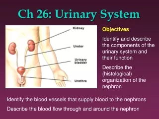
Ch 26: Urinary System Objectives Identify and describe the components of the urinary system and their function Describe the (histological) organization of the nephron Identify the blood vessels that supply blood to the nephrons Describe the blood flow through and around the nephron
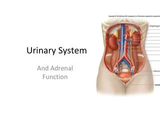
Functions of Urinary System (Kidneys): • Excretion of organic waste products Regulation of blood • Volume • pH • Ion concentration
Kidney Location Lateral to vertebral column in retro-peritoneal position
Surface Anatomy • Hilus Three layers • Renal fascia • Adipose capsule • Renal capsule
Sectional Anatomy Cortex: light reddish brow and granular (due to many capillaries) Medulla: darker striped appearance (due to tubules) Subdivided into distinct renal pyramids. Separated by renal columns from the cortex. Fig 26-3
Urine collection: Ducts within each renal papilla release urine into minor calyx major calyx renal pelvis ureter
Functional unit: Nephron (>mio/kidney) • Renal corpuscle: • Glomerulus • Bowman’s capsule • Tubular passageways: • PCT • LOH • DCT • CD Fig 26-5 Fig 26-4
Filtration: Passage across Three Barriers Capillary endothelium Fenestrated What gets through? Basement membrane Glomerular epithelium (= visceral layer of Bowman’s capsule) slit pores between pedicels Fig 26-5
Juxtaglomerular Apparatus Macula densa + Juxtaglomerular cells (smooth muscle fibers from afferent arteriole) = Juxtaglomerular Apparatus = Endocrine system structure (renin and EPO)
Cortical vs Juxtamedullary Nephrons • Cortical nephrons (85%) • Juxtamedullary nephrons (15%), play keyrole in concentrating urine.
Segmental arteries Interlobar arteries Arcuate arteries Interlobular arteries Afferent arterioles Glomerulus Efferent arterioles Peritubular capillaries Segmental veins Interlobar veins Arcuate veins Interlobular veins Venules Renal Circulation Fig 26-7 Renal Artery Renal Vein
Urine Transport, Storage, and Elimination • Trace drop of urine from kidneys to outside world • Lining of these parts? • Nephroptosis (= floating kidneys) • Nephrolithiasis
Nephroptosis Upright position: 1st degree nephroptosis: Supine position
Nephrolithiasis occurs when urine becomes too concentrated and substances crystalize. Symptoms arise when stones begin to move down ureter causing intense pain. Kidney stones may form in the pelvis or calyces of the kidney or in the ureter.
max. holding capacity: 1l Anatomy of Urinary Bladder Detrusor muscle: • inner longitudinal • middle circular • outer longitudinal
Male versus Female UTIs (esp. E.coli)
The End • Kidneys may sustain 90% loss of nephrons and still not show apparent symptoms!!! • 2-4 % of population only have 1 kidney! Manneken Pis Fountain Brussels, 1619