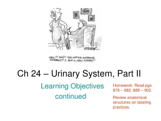
Urinary System Ch 45-47
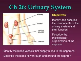
Urinary System Ch 45-47. Zoya Minasyan RN MSN- Edu. Structures and Functions of Urinary System. Assessment of Urinary System. Assessment of Urinary System. Diagnostic Studies of Urinary System. Diagnostic Studies of Urinary System; Cystoscopic examination.

Urinary System Ch 45-47
E N D
Presentation Transcript
Urinary SystemCh 45-47 ZoyaMinasyan RN MSN-Edu
Diagnostic Studies of Urinary System; Cystoscopic examination
Diagnostic study • Ch 45, table 45-8 • Page 1114-1119
Urinary Tract Infection • Bladder and its contents are free of bacteria in most healthy patients. • Escherichia coli most common pathogen • Fungal and parasitic infections can cause UTIs. • Patients at risk • Are immunosuppressed • Have diabetes • Have undergone multiple antibiotic courses • Have traveled to certain Third World countries
Classification • Upper versus lower • Upper tract • Renal parenchyma, pelvis, and ureters • Typically causes fever, chills, flank pain • Example Pyelonephritis: inflammation of renal parenchyma and collecting system • Lower urinary tract • Usually no systemic manifestations • Example Cystitis—Inflammation of bladder wall
Classification • Complicated versus uncomplicated • Uncomplicated • Occurs in otherwise normal urinary tract • Usually involves only the bladder • Complicated • Those with coexisting presence of • Obstruction • Stones • Catheters • Existing diabetes/neurologic disease • Pregnancy-induced changes • Recurrent infection
Classification • Bacterial persistence • Occurs when • Bacteria develop resistance to antibiotic agent. Foreign body in urinary system allows bacteria to survive • Unresolved bacteriuria • Occurs when • Bacteria are resistant to antibiotic • Drug is discontinued before bacteriuria is completely eradicated • Antibiotic agent fails to achieve adequate concentrations in bloodstream or urine to kill bacteria
Etiology and Pathophysiology • Urinary tract above urethra normally sterile • Defense mechanisms exist to maintain sterility/prevent UTIs. • Complete emptying of bladder
Etiology and Pathophysiology • Defense mechanisms • Acidic pH • High urea concentration • Alteration in defense mechanisms increases risk of contracting UTI. • Predisposing factors • Factors increasing urinary stasis • Examples: BPH, tumor, neurogenicbladder,stones • Foreign bodies • Examples: Catheters, instrumentation • Anatomic factors • Examples: Obesity, congenital defects, fistula • Compromising immune response factors • Examples: Age, HIV, diabetes • Functional disorders • Example: Constipation • Other factors • Examples: Pregnancy, multiple sex partners (women)
Etiology and Pathophysiology • Organisms are introduced via the ascending route from the urethra. • Less common routes • Bloodstream • Lymphatic system • Hospital-acquired UTI accounts for 31% of all nosocomial infections. • Causes • Often: E. coli • Seldom: Pseudomonas • Catheter-acquired UTIs • Bacterial biofilms develop on inner surface of catheter.
Clinical Manifestations • Symptoms related to bladder storage or bladder emptying • Bladder storage • Urinary frequency • Abnormally frequent (> every 2 hours) • Urgency • Sudden strong desire to void immediately • Incontinence • Loss or leakage of urine • Nocturia • Waking up ≥2 times at night to void • Nocturnal enuresis • Complaint of loss of urine during sleep • Bladder emptying • Weak stream • Hesitancy • Difficulty starting the urine stream • Intermittency • Interruption of urinary stream while voiding • Postvoid dribbling • Urine loss after completion of voiding • Urinary retention • Inability to empty urine from bladder • Dysuria • Difficulty voiding • Pain on urination • Flank pain, chills, and fever indicate infection of upper tract. • Pyelonephritis
Clinical Manifestations • Older adults • Symptoms are often absent. • Experience nonlocalized abdominal discomfort rather than dysuria • May have cognitive impairment • Are less likely to have a fever • Patients over age 80 years may experience a slight decline in temperature.
Diagnostic Studies • History and physical examination • Dipstick urinalysis • Identify presence of nitrates, WBCs, and leukocyte esterase. • Table 45-8, page 1114
Diagnostic Studies • Urine for culture and sensitivity (if indicated) • Clean-catch sample preferred • Specimen by catheterization or suprapubic needle aspiration more accurate • Determine susceptibility of bacteria to antibiotics • Imaging studies • IVP(IV pyelogram) • Antegradepyelogram • Retrograde pyelogram • Abdominal CT when obstruction suspected • Renal ultrasound for recurrent UTIs
Diagnostic Studies • IVP(IV pyelogram) • Visualizes urinary tract after IV injection of contrast media. Size, shape, position of kidneys, ureters, bladder, tumor, cysts, lesions, obstructions • Nursing • Check for iodine sensitivity • Warmth, a flushed face and salty taste during injection of contrast media • Force fluid after procedure to flush out contrast media. • Antegradepyelogram • If pt has allergy to contrast media or decreased renal fx or no passage to ureteral catheter -contrast media inserted into renal pelvis or via nephrostomy tube • Retrograde pyelogram-X ray • If pt has allergy to contrast, cyctoscope is inserted and ureteral catheter are inserted through it into renal pelvis and contrast is inserted through catheter • Abdominal CT when obstruction suspected • Visualization of kidneys • Masses, tumor • Iv contrast to differentiate masses • KUB- Kidneys, ureters, bladder • Bowel prep if needed • X-ray of abdomen for the size, shape and position of the kidneys • Stones and foreign bodies can be seen • Renal ultrasound for recurrent UTIs • To detect mass
Diagnostic Studies • Teach women • To wipe the periurethral area from front to back using a moistened, clean gauze sponge (no antiseptic is used, as it could contaminate the specimen and cause false-positives) • tell them to collect the specimen 1 to 2 seconds after voiding starts. • Instruct men to wipe the penis around the urethra. The specimen is collected 1 to 2 seconds after voiding begins. • Refrigerate urine immediately on collection
Collaborative Care: Drug Therapy • Trimethoprim/sulfamethoxazole (TMP/SMX) • Used to treat uncomplicated or initial • Inexpensive • Taken twice a day • E. coli resistance to TMP-SMX • Nitrofurantoin (Macrodantin) • Given 3 or 4 times a day • Long-term use • Pulmonary fibrosis • Neuropathies • Fluoroquinolones • Treat complicated UTIs • Example: Ciprofloxacin (Cipro) • Pyridium • Used in combination with antibiotics • Provides soothing effect on urinary tract mucosa • Stains urine reddish orange • Can be mistaken for blood and may stain underclothing • OTC
Nursing Management: Nursing Assessment • Health history • Previous UTIs, calculi, stasis, retention, pregnancy, STDs, bladder cancer • Antibiotics, anticholinergics, antispasmodics • Urinary hygiene • N/V, anorexia, chills, nocturia, frequency, urgency • Suprapubic/lower back pain, bladder spasms, dysuria, burning on urination
Nursing Management: Nursing Assessment • Objective data • Fever • Hematuria, foul-smelling urine, tender, enlarged kidney • Leukocytosis, positive findings for bacteria, WBCs, RBCs, pyuria, ultrasound, CT scan, IVP
Nursing ManagementNursing Diagnoses • Impaired urinary elimination • Ineffective self-health management
Nursing ManagementPlanning • Patient will have • Relief from lower urinary tract symptoms • Prevention of upper urinary tract involvement • Prevention of recurrence
Nursing ManagementNursing Implementation • Health promotion • Recognize individuals at risk. • Debilitated persons • Older adults • Underlying diseases (HIV, diabetes) • Taking immunosuppressive drug or corticosteroids • Emptying bladder regularly and completely • Evacuating bowel regularly • Wiping perineal area front to back • Drinking adequate fluids
Nursing ManagementNursing Implementation • Health promotion (cont’d) • Cranberry juice or cranberry essence may help decrease risk. • Avoid unnecessary catheterization and early removal of indwelling catheters. • Aseptic technique must be followed during instrumentation procedures. • Wash hands before and after contact. • Wear gloves for care of urinary system. • Routine and thorough perineal care for all hospitalized patients • Avoid incontinent episodes by answering call light and offering bedpan at frequent intervals
Nursing Management: Nursing Implementation • Adequate fluid intake • Patient may think will worsen condition because of discomfort. • Dilutes urine, making bladder less irritable • Flushes out bacteria before they can colonize • Avoid caffeine, alcohol, citrus juices, chocolate, and highly spiced foods. • Potential bladder irritants • Emphasize taking full course of prescribed drugs despite disappearance of symptoms. • Second or reduced drug may be ordered after initial course in susceptible patients. • Instruct patient about drug therapy and side effects. Instruct patient to watch urine for changes in color and consistency and decrease in cessation of symptoms. • Application of local heat to suprapubic or lower back may relieve discomfort. • Counsel on persistence of lower tract symptoms beyond treatment; onset of flank pain or fever should be reported immediately
Nursing ManagementNursing Implementation • Ambulatory and home care • Emphasize compliance with drug regimen. • Take as ordered. • Maintain adequate fluids. • Regular voiding (every 3 to 4 hours) • Void after intercourse. • Instruct on follow-up care. • Recurrent symptoms typically occur 1 to 2 weeks after therapy.
Nursing ManagementEvaluation • Use of nonanalgesic relief measures • Appropriate use of analgesics • Passage of urine without urgency • Urine free of blood • Adequate intake of fluids
Acute Pyelonephritis • Inflammation of renal parenchyma(consisting of the nephrones) and collecting system • Caused most commonly by bacteria, Fungi, protozoa, or viruses.
Acute Pyelonephritis Fig. 46-2. Acute pyelonephritis. Cortical surface shows grayish white areas of inflammation and abscess formation (arrow).
Etiology and Pathophysiology • Urosepsis • Systemic infection from urologic source • Can lead to septic shock and death • Septic shock: Outcome of unresolved bacteremia involving gram-negative organism • Usually begins with colonization and infection of lower tract via ascending urethral route • Frequent causes • Escherichia coli • Proteus • Klebsiella • Enterobacter
Collaborative Care • Hospitalization for patients with severe infections and complications • Such as nausea and vomiting with dehydration • Signs/symptoms typically improve within 48 to 72 hours after therapy is started. • The patient with mild symptoms may be treated as an outpatient with antibiotics for 14 to 21 days. • When initial treatment resolves acute symptoms and the patient is able to tolerate oral fluids and drugs, the person may be discharged on a regimen of oral antibiotics for an additional 14 to 21 days.
Nursing Management: Nursing Assessment • Health history • Nausea, vomiting, anorexia, chills, nocturia, frequency, urgency • Suprapubic or lower back pain, bladder spasms, dysuria, burning on urination • Objective data • Fever • Hematuria, foul-smelling urine, tender, enlarged kidney • Leukocytosis, positive findings for bacteria, WBCs, RBCs,ultrasound, CT scan, IVP
Nursing Management • Nursing Diagnoses • Acute pain • Impaired urinary elimination • Planning • Patient will have • Relief of pain • Normal body temperature • No complications • Normal renal function • No recurrence of symptoms
Nursing ManagementNursing Implementation • Health promotion • Early treatment for cystitis to prevent ascending infection • Patient with structural abnormalities is at high risk • Stress the need for regular medical care. • Ambulatory and home care • Need to continue drugs as prescribed • Need for follow-up urine culture • Identification of risk for recurrence • Encourage adequate fluids. • Rest to increase comfort • Low-dose, long-term antibiotics to prevent re infections
Nursing ManagementEvaluation • Appropriate use of analgesics • Passage of urine without urgency • Urine free of blood • Adequate intake of fluids
Question The nurse identifies the patient with the greatest risk for a urinary tract infection as a: 1. 37-year-old man with kidney stones. 2. 26-year-old pregnant woman who has a history of urinary tract infection. 3. 69-year-old man who has urinary retention caused by benign prostatic hyperplasia. 4. 72-year-old woman hospitalized with a stroke who has a urinary catheter because of urinary incontinence.
Chronic Kidney Disease (CKD) • Involves progressive, irreversible loss of kidney function • Disease staging based on decrease in GFR • Normal GFR 125 mL/min, which is reflected by urine creatinine clearance • Last stage of kidney failure • End-stage renal disease (ESRD) occurs when GFR <15 mL/min
Chronic Kidney Disease • Defined as presence of • Kidney damage • Pathologic abnormalities • Markers of damage • Blood, urine, imaging tests • Glomerular filtration rate (GFR) • <60 mL/min for 3 months or longer
Chronic Kidney Disease • Leading causes of ESRD • Diabetes • Hypertension
Clinical ManifestationsUrinary System • Polyuria • Results from inability of kidneys to concentrate urine • Occurs most often at night • Oliguria • Occurs as CKD worsens • 300-500 ml/day • Anuria • Urine output <40 mL per 24 hours
Clinical ManifestationsMetabolic Disturbances • Waste product accumulation • As GFR ↓, BUN ↑ and serum creatinine levels ↑ • BUN ↑ • Not only by kidney failure but by protein intake, fever, corticosteroids, and catabolism • N/V, lethargy, fatigue, impaired thought processes, and headache may occur. • Altered carbohydrate metabolism • Caused by impaired glucose use • From cellular insensitivity to the normal action of insulin • Defective carbohydrate metabolism • Patients with diabetes who become uremic may require less insulin than before onset of CKD. • Insulin dependent on kidneys for excretion
Clinical Manifestations Metabolic Disturbances • Elevated triglycerides • Hyperinsulinemia stimulates hepatic production of triglycerides. • Altered lipid metabolism • ↓ levels of enzyme lipoprotein lipase • Important in breakdown of lipoproteins
Clinical ManifestationsElectrolyte/Acid-Base Imbalances • Potassium • Hyperkalemia • Most serious electrolyte disorder in kidney disease • Fatal dysrhythmias • Sodium • May be normal or low • Because of impaired excretion, sodium is retained. • Water is retained. • Edema • Hypertension • CHF • Calcium and phosphate alterations • Magnesium alterations
Clinical Manifestations Electrolyte/Acid-Base Imbalances • Metabolic acidosis • Results from • Inability of kidneys to excrete acid load (primary ammonia) • Defective reabsorption/regeneration of bicarbonate • The average adult produces 80 to 90 mEq of acid per day. This acid is normally buffered by bicarbonate. • In kidney failure, plasma bicarbonate, which is an indirect measure of acidosis, usually falls to a new steady state at around 16 to 20 mEq/L (16 to 20 mmol/L).