Glycosphingolipid Function in Atherosclerosis
780 likes | 865 Vues
Discover the structure, function, and methods for determining glycosphingolipids' role in atherosclerosis and vascular biology. Learn about lysosomal storage disorders and the prevalence of glycosphingolipid disorders.

Glycosphingolipid Function in Atherosclerosis
E N D
Presentation Transcript
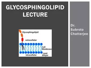
GLYCOSPHINGOLIPID LECTURE Dr. SubrotoChatterjee
What are glycosphingolipids? The most common characteristic component of glycosphingolipids is the aliphatic amino alcohol discovered by Thudichum. Thudichum named it Sphingosine after the enigmatic Sphinx from Egypt having a head of Pharaoh and body of a lion.
Lysosomal storage disorders • Several metabolic basis of inherited diseases in man occur due to the lack/deficiency of enzymes which catabolize glycosphingolipids
Glycosphingolipid lecture topics • Function • Methods for Determining Function • Extraction and Purification • Quantitation • Structural Determination • Localization • Imaging • Metabolism
Function of glycosphingolipids • Superoxide generation and CAM expression • Inhibition of nitric oxide production in endothelial cells • As mediators of growth factors contributing to cell proliferation • As receptors for toxins and bacteria
Function of glycosphingolipids in atherosclerosis and vascular Biology • 1) Generation of superoxide in arterial smooth muscle cells and expression of cell adhesion molecules.
Lactosylceramide mediates tnfα-induced icam-1 expression in endothelial cells
Lactosylceramide stimulates superoxide generation in human endothelial cells
Lactosylceramide stimulates superoxide generation in human endothelial cells
Function of glycosphingolipids in atherosclerosis and vascular Biology • 2) Inhibition of nitric oxide production in endothelial cells.
Effect of laccer on endothelium dependent vaso-relaxation and porcine coronary artery
Function of glycosphingolipids in atherosclerosis and vascular Biology • 3. As mediators of growth factors contributing to cell proliferation and angiogenesis
Function of glycosphingolipids • 5) Serve as receptors to various toxins, e.g. cholera toxin and other bacteria
Methods FOR determining gsl function • Cellular assays Proliferation • Adhesion Angiogenesis • Migration • Apoptosis
Gsl extraction • 1) Bligh and Dyer • 2) Folch Partitioning
Extraction of Glycosphingolipids from Heart Tissue • Modified Bligh & Dyer Method • extraction in chloroform:methanol 2:1 • homogenized by hand • lipids extracted from organic phase https://encrypted-tbn0.gstatic.com/images?q=tbn:ANd9GcSoXJZC7o-xxP_UJqYeqM-GpLvFH_2Vq-18XdegMaMqdjDx9_V2 http://www.sfu.ca/bisc/bisc-429/folch.gif Analysis of Glycosphingolipids Subroto Chatterjee October 7, 2013
Purification glycosphingolipids • Alkaline methanolysis • The Glycolipid fraction from the silicic acid column is treated with mild base to remove contaminating phospholipids. This treatment does not affect glycolipids or gangliosides unless they contain an O-acyl group. The following quantities are used for 1-10mg of glycolipid fraction. Add 1ml of chloroform and 1ml of 0.6 NNaOH in methanol to the dry fraction and allow the mixture to react at room temperature for 1 hour. Then add 1.2 ml of 0.5 N HCL in methanol, 1.7ml of water, and 3.4ml of chloroform, mix well, centrifuge, and remove the lower layer containing the glycolipids. Was the lower layer three times with methanol:water (1:1) and then evaporate it to dryness in vacuo.
Hptlcanalysis of neutral gsl • Mixture of compounds in lane C through F • Monohexosylceramide, gal- and glc-ceramide • Dihexosylceramide, gal(1->4) glc-ceramide and gal(1->4)gal-ceramide • Trihexosylceramide, gal(1->4)gal(1->4)glc-cermaide • TetrahexosylceramidegalNAc(1->3)gal(1->4)gal(1->4)glc-ceramide • On a silica gel H plate developed with chloroform-methanol-water (100:42:6) and visualized with alpha-napthol spray
Hptlc analysis of gangliosides Thin-layer chromatogram disaloganglioside, NANA (2->3)gal(1->3)galNAc(14)[NANA(2->3)]gal(1->4)glc-ceramide Monosialoganglioside, galNAc(1->4)[NANA(2->3)gal(1->4)glc-ceramide On a silica gel G (0.25mm) plate developed two times with chloroform-methanol-2.5N NH4OH (65:45:9) and visualized with alpha-naphtol spray
Quantitation of Glycosphingolipids • Gas chromatography of sugars • TMSI derivatization • MS analysis • TMSI derivatization • HPLC analysis • perbenzoylation(McCluer) • endoglycoceramidation(Butters) • HPTLC and densitometricscanning • HPLC analysis • deacylation (Zama) • MS analysis • Without derivatization
Quantitation of Glycosphingolipids • Gas chromatography of sugars • TMSI derivatization • MS analysis • TMSI derivatization • HPLC analysis • perbenzoylation (McCluer) • endoglycoceramidation (Butters) • HPTLC and densitometric scanning • HPLC analysis • deacylation (Zama) • MS analysis • Without derivatization
Trimethylsilylation and gas-liquid chromatography of methyl glycosides
Quantitation of Glycosphingolipids • Gas chromatography of sugars • TMSI derivatization • MS analysis • TMSI derivatization • HPLC analysis • perbenzoylation(McCluer) • endoglycoceramidation(Butters) • HPTLC and densitometricscanning • HPLC analysis • deacylation (Zama) • MS analysis • Without derivatization
Mass spectrometry of intact tmsi derivatives of glycolipids • Mass spectrometry of intact TMS derivatives of glycolipids gives information about the sugar groups, the fatty acid and the sphingosine portion of glycosphingolipids. Bis (trimethylsiyl) trifluroroacetamide (100microliter) and pyridine (50 microliter) are added to 20-200microgram of the purified glycospingolipid in a small capped vial and heated at 60 degrees F for about 30 minutes. An aliquot containing 10-20 microgram of the TMS glycolipid is evaporated to dryness under nitrogen in a mass spectrometer direct probe tube. The samples are volatilized in the mass spectrometer ion source at temperatures ranging 100 degrees to 180 degrees depending on the size of the oligosaccharide unit. • The following information can be obtained by comparison of the resulting mass spectra with those of reference samples: (1) whether the terminal residue is a hexose or hexosamine; (2) the number of and nature of N-acetylneuramine acid groups (i.e., terminal or branched); (3) whether N-acetyl and/or N-glycolylneuraminate is present; (4) information regarding the number of glycosyl residues present and the fatty acid and sphingosine composition. It is essential, because of the limitations of this technique (e.g., the inability to distinguish between hexoses), that it be used in conjunction with other techniques, such as permethylation analyses, and studies with specific glycosidases.
Quantitation of Glycosphingolipids • Gas chromatography of sugars • TMSI derivatization • MS analysis • TMSI derivatization • HPLC analysis • perbenzoylation(McCluer) • endoglycoceramidation(Butters) • HPTLC and densitometricscanning • HPLC analysis • deacylation (Zama) • MS analysis • Without derivatization
Perbenzoylation McCluer: Methods in Enzymology 138: 1987 • Principle: • Since neutral glycosphingolpids do not possess a characteristic chromophore that permits their quantitation by UV detection, they can be derivatized with benzoylchloride to form stable per-O,N benzoylated products. These products can be quantified by UV light at 280nm or at a higher sensitivity at 230nm. • GSL(200ng) plus N-acetylpsychosine (an internal standard) are dried in N2. Perbenzoylation is carried out by adding 500uL of 10% benzoylchloride in pyridine for 16hr at 37 C. The samples are N2 dried and washed thrice with 1.8mL of methanol: water(saturated with sodium carbonate). The hexane layer is washed and finally dried in N2. The derivatives are dissolved in CCL4(100uL) and a suitable aliquot injected in to the HPLC column(Zipax,E.I DuPont column 2.1 mmx500nm). HPLC of male (C57BL/6J) mouse kidney perbenzoylated glycosphingolipids on a Zipax column with detection at 230nm
Quantitation of Glycosphingolipids • Gas chromatography of sugars • TMSI derivatization • MS analysis • TMSI derivatization • HPLC analysis • perbenzoylation(McCluer) • endoglycoceramidation(Butters) • HPTLC and densitometricscanning • HPLC analysis • deacylation (Zama) • MS analysis • Without derivatization
Quantitation of Glycosphingolipids • Gas chromatography of sugars • TMSI derivatization • MS analysis • TMSI derivatization • HPLC analysis • perbenzoylation(McCluer) • endoglycoceramidation(Butters) • HPTLC and densitometricscanning • HPLC analysis • deacylation (Zama) • MS analysis • Without derivatization
Quantitation of Glycosphingolipids • 3) Endoglycoceramidaseuse to excise the oligosaccharides for quantification by HPLC. • Wing, D. R., et al. "High-performance liquid chromatography analysis of ganglioside carbohydrates at the picomole level after ceramideglycanase digestion and fluorescent labeling with 2-aminobenzamide." Analytical biochemistry 298.2 (2001): 207-217.
Increased glycosphingolipid levels in serum and aortaeof apolipoprotein E gene knockout mice
Quantitation of Glycosphingolipids • Gas chromatography of sugars • TMSI derivatization • MS analysis • TMSI derivatization • HPLC analysis • perbenzoylation(McCluer) • endoglycoceramidation(Butters) • HPTLC and densitometricscanning • HPLC analysis • deacylation (Zama) • MS analysis • Without derivatization
Quantitation of Glycosphingolipids • Gas chromatography of sugars • TMSI derivatization • MS analysis • TMSI derivatization • HPLC analysis • perbenzoylation(McCluer) • endoglycoceramidation(Butters) • HPTLC and densitometricscanning • HPLC analysis • deacylation (Zama) • MS analysis • Without derivatization
Quantitation of Glycosphingolipids using deacylase • 2) deacylase treatment and quantification of lysoGSL • Lipid Extraction • Modified Bligh and Dyer • N-deacylation • Dried sphingolipid standards and samples were deacylated using sphingolipidceramide N-deacylase (SCDase). To each dried sample and standard, 27uL 25mM sodium acetate buffer (pH 5.5) was added • To this solution, 5uL SCDase was added • Samples and standards were enzymatically digested for 19h • The reaction was stopped with 200uL chloroform-methanol (1:1, v/v) • The organic layer was removed and dried under N2 gas. • Derivatization • Samples were derivatized with 15uL OPA solution. • HPLC • Agilent 1260 Infinity using a Zorbax SB-C18 reversed-phase column • Solvent System: isocratic (methanol:water acidified with 0.2% trifluoroacetic acid at 88:12, v/v) • Flow Rate: 0.75 mL/min • Ex λ: 340nm Emλ: 360nm
Quantitation of Glycosphingolipids • Gas chromatography of sugars • TMSI derivatization • MS analysis • TMSI derivatization • HPLC analysis • perbenzoylation(McCluer) • endoglycoceramidation(Butters) • HPTLC and densitometricscanning • HPLC analysis • deacylation (Zama) • MS analysis • Without derivatization