Download

1 / 27
270 likes | 416 Vues
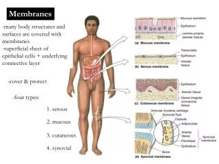
Diagrams For Chapter 4 Test Skin & Body Membranes. Anatomy & Physiology Honors Instructor: Phyllis Smith Jefferson Township High School. Figure 4.1a Classes of epithelial membranes. (a) Cutaneous membrane. Cutaneous membrane (skin). (a) Cutaneous membrane.

E N D
Diagrams For Chapter 4 TestSkin & Body Membranes • Anatomy & Physiology Honors • Instructor: Phyllis Smith • Jefferson Township High School
Figure 4.1a Classes of epithelial membranes. (a) Cutaneous membrane. Cutaneous membrane (skin) (a) Cutaneous membrane
Figure 4.1b Classes of epithelial membranes. (b) Mucous membranes. Mucosa of nasal cavity Mucosa of mouth Esophagus lining Mucosa of lung bronchi (b) Mucous membranes
Figure 4.1c Classes of epithelial membranes. (c) Serous membranes. Parietal peritoneum Visceral peritoneum (c) Serous membranes
Figure 4.1d Classes of epithelial membranes. (d) Relationship between the parietal and visceral serous membrane layers. Parietal pleura Visceral pleura Parietal pericardium Visceral pericardium Outer wall (comparable to parietal serosa) Air or water (comparable to serous fluid) Inner wall (comparable to visceral serosa) (d)
Figure 4.2 A typical synovial joint. Ligament Joint cavity (contains synovial fluid) Articular (hyaline) cartilage Fibrous capsule Articular capsule Synovial membrane
Figure 4.3 The epidermis of thick skin. Epidermis • Stratum corneum • Stratum lucidum • Stratum granulosum • Stratum spinosum • Stratum basale Dermis
Skin structure Sensory nerve fiber Hair follicle receptor
Figure 4.4 Skin structure. Hair shaft Pore Dermal papillae Epidermis Arrector pili muscle Papillary layer Appendages of the skin: Sebaceous (oil) gland Reticular layer Dermis Eccrine sweat gland Hair follicle Hair root Hypodermis (subcutan- eous tissue) Adipose (fat) tissue Nervous structures: Meissner’s corpuscle Cutaneous blood vessels Pacinian corpuscle Sensory nerve fiber Hair follicle receptor
Appendages of the Skin Figure 4.6b
Appendages of the Skin Figure 4.6a
Figure 4.7a Structure of a hair and hair follicle. (a) Longitudinal section of a hair within its follicle. (a)
Figure 4.7a Structure of a hair and hair follicle. (a) Longitudinal section of a hair within its follicle. Hair shaft Arrector pili Sebaceous gland Hair root Hair bulb in follicle (a)
Figure 4.7b Structure of a hair and hair follicle. (b) Hair. (b) Hair
Figure 4.7b Structure of a hair and hair follicle. (b) Hair. Cuticle Cortex Medulla (b) Hair
Figure 4.7c Structure of a hair and hair follicle. (c) Enlarged longitudinal view of the expanded hair bulb in the follicle showing the matrix, the region of actively dividing epithelial cells that produces the hair. (c)
Figure 4.7c Structure of a hair and hair follicle. (c) Enlarged longitudinal view of the expanded hair bulb in the follicle showing the matrix, the region of actively dividing epithelial cells that produces the hair. Dermal sheath Hair follicle Epidermal sheath Matrix (growth zone) in hair bulb Melanocyte Connective tissue papilla containing blood vessels (c)
Figure 4.9a Structure of a nail. (a) Surface view. Surface view
Figure 4.9a Structure of a nail. (a) Surface view. Body of nail Lateral nail fold Lunula Cuticle (a) Surface view
Figure 4.9b Structure of a nail. (b) Longitudinal section of the distal part of a finger. (b) Longitudinal section of the distal part of a finger
Figure 4.9b Structure of a nail. (b) Longitudinal section of the distal part of a finger. Nail matrix Root of nail Proximal nail fold Nail bed Free edge of nail Cuticle Body of nail Bone of fingertip Stratum basale (b) Longitudinal section of the distal part of a finger