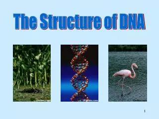
Solving the Structure of DNA
110 likes | 130 Vues
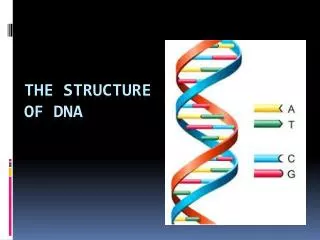
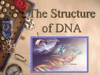
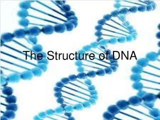
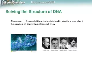
Solving the Structure of DNA. The research of several different scientists lead to what is known about the structure of deoxyribonucleic acid, DNA. Chargaff’s Rules. Erwin Chargaff discovered that the percentages of adenine [A] and thymine [T] bases are almost equal in any sample of DNA.

Solving the Structure of DNA
E N D
Presentation Transcript
Solving the Structure of DNA • The research of several different scientists lead to what is known about the structure of deoxyribonucleic acid, DNA.
Chargaff’s Rules • Erwin Chargaff discovered that the percentages of adenine [A] and thymine [T] bases are almost equal in any sample of DNA. • The same thing is true for the other two nucleotides, guanine [G] and cytosine [C]. • The observation that [A] = [T] and [G] = [C] became known as one of “Chargaff’s rules.”
Franklin’s X-Rays • In the 1950s, British scientist Rosalind Franklin used a technique called X-ray diffraction to get information about the structure of the DNA molecule.
Franklin’s X-Rays • X-ray diffraction revealed an X-shaped pattern showing that the strands in DNA are twisted around each other like the coils of a spring. • The angle of the X-shaped pattern suggested that there are two strands in the structure. • Other clues suggest that the nitrogenous bases are near the center of the DNA molecule.
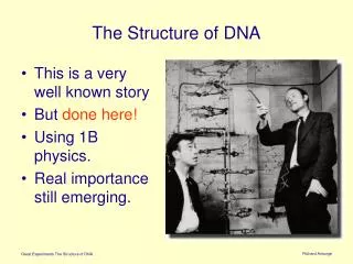
The Work of Maurice Wilkins • Wilkins studied biological molecules like DNA and viruses using a variety of microscopes and spectrophotometers. He eventually began using X-rays to produce diffraction images of DNA molecules. The X-ray diffraction images produced by him, Rosalind Franklin, and Raymond Gosling led to the deduction by James Watson and Francis Crick of the 3-dimensional helical nature of DNA. Wilkins shared the 1962 Nobel Prize in Physiology or Medicine with Watson and Crick.
The Work of Watson and Crick • At the same time, James Watson, an American biologist, and Francis Crick, a British physicist, were also trying to understand the structure of DNA. • They built three-dimensional models of the molecule.
The Work of Watson and Crick • Early in 1953, without her permission, Wilkins showed Watson a copy of Franklin’s X-ray pattern. • The clues in Franklin’s X-ray pattern enabled Watson and Crick to build a model that explained the specific structure and properties of DNA. • Watson and Crick’s breakthrough model of DNA was a double helix, in which two strands were wound around each other.
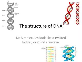
The Double-Helix Model • A double helix looks like a twisted ladder. • In the double-helix model of DNA, the two strands twist around each other like spiral staircases. • The double helix accounted for Franklin’s X-ray pattern and explains Chargaff’s rule of base pairing and how the two strands of DNA are held together.
Hydrogen Bonding • Watson and Crick discovered that hydrogen bonds could form between certain nitrogenous bases, providing just enough force to hold the two DNA strands together. • Hydrogen bonds are relatively weak chemical forces that allow the two strands of the helix to separate. • The ability of the two strands to separate is critical to DNA’s functions.
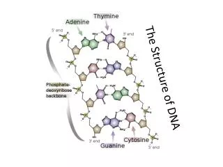
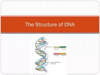
Base Pairing • Watson and Crick’s model showed that hydrogen bonds could create a nearly perfect fit between nitrogenous bases along the center of the molecule. • These bonds would form only between certain base pairs—adenine with thymine, and guanine with cytosine. • This nearly perfect fit between A–T and G–C nucleotides is known as base pairing, and is illustrated in the figure.
Base Pairing • Watson and Crick realized that base pairing explained Chargaff’s rule. It gave a reason why [A] = [T] and [G] = [C]. • For every adenine in a double-stranded DNA molecule, there had to be exactly one thymine. For each cytosine, there was one guanine.