
DIGESTIVE SYSTEM
DIGESTIVE SYSTEM. DIGESTIVE SYSTEM. The digestive system is the collective name used to describe the alimentary canal, some accessory organs and a variety of digestive processes which take place at different levels in the canal to prepare food eaten in the diet for absorption.

DIGESTIVE SYSTEM
E N D
Presentation Transcript
DIGESTIVE SYSTEM Meghna.D.Punjabi
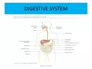
DIGESTIVE SYSTEM • The digestive system is the collective name used to describe the alimentary canal, some accessory organs and a variety of digestive processes which take place at different levels in the canal to prepare food eaten in the diet for absorption. • The alimentary canal begins at the mouth, passes through the thorax, abdomen and pelvis and ends at the anus. Meghna.D.Punjabi
ACTIVITIES OF DIGESTIVE SYSTEM The activities in the digestive system can be grouped under five main headings. • Ingestion. This is the process of taking food into the alimentary tract. • Propulsion. This moves the contents along the alimentary tract. • Digestion. • This consists of mechanical breakdown of food by, e.g. mastication (chewing) • chemical digestion of food by enzymes present in secretions produced by glands and accessory organs of the digestive system. • Absorption. • This 'is the process by which digested food substances pass through the walls of some organs of the alimentary canal into the blood and lymph capillaries for circulation around the body. • Elimination. • Food substances which have been eaten but cannot be digested and absorbed are excreted by the bowel as faeces. Meghna.D.Punjabi
ORGANS OF THE DIGESTIVE SYSTEM Alimentary tract • This is a long tube through which food passes. It commences at the mouth and terminates at the anus, and the various parts are given separate names, although structurally they are remarkably similar. The parts are: • mouth • pharynx • oesophagus • stomach • small intestine • large intestine • rectum and anal canal. Meghna.D.Punjabi
ACCESSORY ORGANS Accessory organs • Various secretions are poured into the alimentary tract, some by glands in the lining membrane of the organs, e.g. gastric juice secreted by glands in the lining of the stomach, and some by glands situated outside the tract. The latter are the accessory organs of digestion and their secretions pass through ducts to enter the tract. They consist of: • 3 pairs of salivary glands • pancreas • liver and the biliary tract. Meghna.D.Punjabi
BASIC STRUCTURE OF THE ALIMENTARY CANAL • The layers of the walls of the alimentary canal follow a consistent pattern from the oesophagus onwards. • This basic structure does not apply so obviously to the mouth and the pharynx. • In the different organs from the oesophagus onwards, modifications of structure are found which are associated with special functions. Meghna.D.Punjabi
ALIMENTARY CANAL Meghna.D.Punjabi
WALLS OF ALIMENTARY TRACT The walls of the alimentary tract are formed by four layers of tissue: • adventitia or outer covering • muscle layer • submucosal layer • mucosa -lining Meghna.D.Punjabi
PERITONEUM Meghna.D.Punjabi
ADVENTIA (OUTER COVERING) • In the thorax this consists of loose fibrous tissue and in the abdomen the organs are covered by a serous membrane called peritoneum . Peritoneum • The peritoneum is the largest serous membrane of the body . • It consists of a closed sac, containing a small amount of serous fluid, within the abdominal cavity. • It is richly supplied with blood and lymph vessels, and contains a considerable number of lymph nodes. • It provides a physical barrier to local spread of infection, and can isolate an infective focus such as appendicitis, preventing involvement of other abdominal structures. It has two layers PARIETAL LAYER: Lines abdominal wall. VISCERAL LAYER: Covers the organs (viscera) within the abdominal and pelvic cavities. • The 2 layers of peritoneum are actually in contact, and friction between them is prevented by the presence of serous fluid secreted by peritoneal cells, thus the peritoneal cavity is called potential cavity. Meghna.D.Punjabi
PERISTALSIS Meghna.D.Punjabi
MUSCLE LAYER • This consists of two layers of smooth (Involuntary) muscle. • The muscle fibres of the outer layer are arranged longitudinally, and those of the inner layer encircle the wall of the tube. Between these two muscle layers are blood vessels, lymph vessels and a plexus (network) of sympathetic and parasympathetic nerves, called the myentericor Auerbach's plexus. These nerves supply the adjacent smooth muscle and blood vessels. • Contraction and relaxation of these muscle layers occurs in waves which push the contents of the tract onwards. This type of contraction of smooth muscle is called peristalsis . • Muscle contraction also mixes food with the digestive juices. Onward movement of the contents of the tract is controlled at various points by sphincters consisting of an increased number of circular muscle fibres. They also act as valves preventing backflow in the tract. The control allows time for digestion and absorption to take place. Meghna.D.Punjabi
SUBMUCOSA LAYER • This layer consists of loose connective tissue with some elastic fibres. • Within this layer are· plexuses of blood vessels and nerves, lymph vessels and varying amounts of lymphoid tissues. The blood vessels consist of arterioles, venules and capillaries. • The nerve plexus is the submucosalor Meissner's plexus, consisting of sympathetic and parasympathetic nerves which supply the mucosal lining. Meghna.D.Punjabi
MUCOSA This consists of three layers of tissue: • mucous membrane formed by columnar epithelium is the innermost layer and has three main functions: protection, secretion and absorption. • lamina propriaconsisting of loose connective tissue, which supports the blood vessels that nourish the inner epithelial layer, and varying amounts of lymphoid tissue that has a protective function. • muscularis mucosa, a thin outer layer of smooth muscle that provides involutions of the mucosa layer, e.g. gastric glands, villi Meghna.D.Punjabi
COLUMNAR EPITHELIUM Meghna.D.Punjabi
MUCOUS MEMBRANE • In parts of the tract which are subject to great wear and tear or mechanical injury this layer consists of stratified squamous epithelium with mucus-secreting glands just below the surface. • In areas where the food is already soft and moist and where secretion of digestive juices and absorption occur, the mucous membrane consists of columnar epithelial cells interspersed with mucus-secreting goblet cells . • Mucus lubricates the walls of the tract and protects them from digestive enzymes. Below the surface in the regions lined with columnar epithelium are collections of specialised cells, or glands, which pour their secretions into the lumen of the tract. These secretions are digestive juices and they contain the enzymes which chemically break down food include: • saliva from the salivary glands • gastric juice from the gastric glands • intestinal juice from the intestinal glands • pancreatic juice from the pancreas • bile from the liver. Meghna.D.Punjabi
MOUTH Meghna.D.Punjabi
MOUTH • The oral cavity is lined throughout with mucous membrane, consisting of stratified squamous epithelium containing small mucus-secreting glands. • The part of the mouth between the gums (alveolar ridges) and the cheeks is the vestibule and the remainder of the cavity is the mouth proper. • The mucous membrane lining of the cheeks and the lips is reflected on to the gums or alveolar ridges and is continuous with the skin of the face. • The palate forms the roof of the mouth and is divided into the anterior hard palate and the posterior soft palate. Meghna.D.Punjabi
MOUTH • The bones forming the hard palate are the maxilla and the palatine bones. The soft palate is muscular, curves downwards from the posterior end of the hard palate and blends with the walls of the pharynx at the sides. • The uvula is a curved fold of muscle covered with mucous membrane, hanging down from the middle of the free border of the soft palate. • Originating from the upper end of the uvula there are four folds of mucous membrane, two passing downwards at each side to form membranous arches. • The posterior folds, one on each side, are the palatopharyngeal arches and the two anterior folds are the palatoglossal arches. On each side, between the arches, is a collection of lymphoid tissue called the palatine tonsil. Meghna.D.Punjabi
TONGUE Meghna.D.Punjabi
PAPILAE OF TONGUE Meghna.D.Punjabi
TONGUE • The tongue is a voluntary muscular structure which occupies the floor of the mouth. It is attached by its base to the hyoid bone and by a fold of its mucous membrane covering, called the frenulum, to the floor of the mouth. The superior surface consists of stratified squamous epithelium, with numerous papillae (little projections), containing nerve endings of the sense of taste, sometimes called the taste buds. There are three varieties of papillae . • Vallate papillae, usually between 8 and 12 altogether, are arranged in an inverted V shape towards the base of the tongue. These are the largest of the papillae and are the most easily seen. • Fungiform papillae are situated mainly at the tip and the edges of the tongue and are more numerous than the vallate papillae. • Filiform papillae are the smallest of the three types. They are most numerous on the surface of the anterior two-thirds of the tongue. Meghna.D.Punjabi
FUNCTIONS OF TONGUE Functions of the tongue • The tongue plays an important part in: • mastication (chewing) • deglutition (swallowing) • speech • taste . • Nerve endings of the sense of taste are present in the papillae and widely distributed in the epithelium of the tongue, soft palate, pharynx and epiglottis. Meghna.D.Punjabi
PERMENANT TEETH Meghna.D.Punjabi
TEETH • The teeth are embedded in the alveoli or sockets of the alveolar ridges of the mandible and the maxilla . Each individual has two sets, or dentitions, the temporary or deciduous teeth and Permanent teeth. At birth the teeth of both dentitions an present in immature form in the mandible and maxilla. • There are 20 temporary teeth, 10 in each jaw. They begin to erupt when the child is about 6 months old, and should all be present after 24 months. • The permanent teeth begin to replace the deciduous teeth in the 6th year of age and this dentition, consisting of 32 teeth, is usually complete by the 24th year. Meghna.D.Punjabi
PERMENANT & DECIDUOUS Meghna.D.Punjabi
PERMENANT & DECIDOUS Meghna.D.Punjabi
PERMENANT TEETH Meghna.D.Punjabi
FUNCTIONS OF TEETH • The incisor and canine teeth are the cutting teeth and are used for biting off pieces of food, whereas the premolar and molar teeth, with broad, flat surfaces, are used for grinding or chewing food. Meghna.D.Punjabi
STRUCTURE OF A TOOTH Meghna.D.Punjabi
STRUCTURE OF A TOOTH • Although the shapes of the different teeth vary, the structure is the same and consists of: • the crown - the part which protrudes from the gum • the root-the part embedded in the bone • the neck-the slightly narrowed region where the crown merges with the root • In the centre of the tooth is the pulp cavity containing blood vessels, lymph vessels and nerves, and surrounding this is a hard ivory-like substance called dentine. Outside the dentine of the crown is a thin layer of very hard substance, the enamel. The root of the tooth, on the other hand, is covered with a substance resembling bone, called cement, which fixes the tooth in its socket. • Blood vessels and nerves pass to the tooth through a small foramen at the apex of each root Meghna.D.Punjabi
SALIVARY GLANDS Meghna.D.Punjabi
SALIVARY GLANDS • Salivary glands pour their secretions into the mouth. There are three pairs: the parotid glands, the submandibular glands and the sublingual glands. Parotid glands • These are situated one on each side of the face just below the external acoustic meatus . Each gland has a parotid duct opening into the mouth at the level of the second upper molar tooth. Submandibular glands • These lie one on each side of the face under the angle of the jaw. The two submandibular ducts open on the floor of the mouth, one on each side of the frenulum of the tongue. Sublingual glands • These glands lie under the mucous membrane of the floor of the mouth in front of the submandibular glands. They have numerous small ducts that open into the floor of the mouth. Meghna.D.Punjabi
COMPOSITION OF SALIVA • Saliva is the combined secretions from the salivary glands and the small mucus-secreting glands of the lining of the oral cavity. About 1.5 litres of saliva is produced daily and it consists of • water • mineral salts • enzyme: salivary amylase • mucus • lysozyme • immunoglobulins • blood-clotting factors. Meghna.D.Punjabi
SECRETION OF SALIVA • Secretion of saliva is under autonomic nerve control. • Parasympathetic stimulation causes vasodilatation and profuse secretion of watery saliva with a relatively low content of enzymes and other organic substances. • Sympathetic stimulation causes vasoconstriction and secretion of small amounts of saliva rich in organic material, especially from the submandibular glands. • Reflex secretion occurs when there is food in the mouth and the reflex can easily become conditioned so that the sight, smell and even the thought of food stimulates the flow of saliva. Meghna.D.Punjabi
STRUCTURE OF SALIVARY GLANDS • The glands are all surrounded by a fibrous capsule. • They consist of a number of lobules made up of small acini lined with secretory cells . • The secretions are poured into ductules which join up to form larger ducts leading into the mouth Meghna.D.Punjabi
FUNCTIONS OF SALIVA Chemical digestion of polysaccharides. • Saliva contains the enzyme amylase that begins the breakdown of complex sugars, reducing them to the disaccharide maltose. The optimum pH for the action of salivary amylase is 6.8 (slightly acid). Salivary pH ranges from 5.8 to 7.4 depending on the rate of flow; the higher the flow rate, the higher is the pH. Enzyme action continues during swallowing until terminated by the strongly acidic pH (1.5 to 1.8) of the gastric juices, which degrades the amylase. Lubrication of food. • Dry food entering the mouth is moistened and lubricated by saliva before it can be made into a bolus ready for swallowing. Meghna.D.Punjabi
FUNCTIONS OF SALIVA Cleansing and lubricating. • An adequate flow of saliva is necessary to cleanse the mouth and keep its tissues soft, moist and pliable. It helps to prevent damage to the mucous membrane by rough or abrasive foodstuffs. Non-specific defence. • Lysozyme, immunoglobulins and clotting factors combat invading microbes. Taste. • The taste buds are stimulated only by chemical substances in solution. Dry foods stimulate the sense of taste only after thorough mixing with saliva. The senses of taste and smell are closely linked in the enjoyment, or otherwise, of food. Meghna.D.Punjabi
PHARYNX • The pharynx is divided for descriptive purpose into three parts, the nasopharynx, oropharynx and laryngopharynx • . The nasopharynx is important in respiration. • The oropharynx and laryngopharynx are passages common to both the respiratory and the digestive systems. • Food passes from the oral cavity into the pharynx then to the oesophagus below, with which it is continuous. Meghna.D.Punjabi
PHARYNX • The walls of the pharynx are built of three layers of tissue. • The lining membrane (mucosa) is stratified squamous epithelium, continuous with the lining of the mouth at one end and with the oesophagus at the other. • The middle layer consists of fibrous tissue which becomes thinner towards the lower end and contains blood and lymph vessels and nerves. • The outer layer consists of a number of involuntary constrictor muscles which are involved in swallowing. When food reaches the pharynx swallowing is no longer under voluntary control. Meghna.D.Punjabi
OESOPHAGUS Meghna.D.Punjabi
OESOPHAGUS • The oesophagus is about 25 cm long and about 2 cm in diameter and lies in the median plane in the thorax in front of the vertebral column behind the trachea and the heart. • It is continuous with the pharynx above and just below the diaphragm it joins the stomach. • Immediately the oesophagus has passed through the diaphragm it curves upwards before opening into the stomach. This sharp angle is believed to be one of the factors which prevents the regurgitation (backward flow) of gastric contents into the oesophagus. Meghna.D.Punjabi
OESOPHAGUS • The upper and lower ends of the oesophagus are closed by sphincter muscles. • The upper cricopharyngeal sphincter prevents air passing into the oesophagus during inspiration and the aspiration of oesophageal contents. • The cardiac or lower oesophageal sphincter prevents the reflux of acid gastric contents into the oesophagus. Meghna.D.Punjabi
STRUCTURE OF OESOPHAGUS There are four layers • . As the oesophagus is almost entirely in the thorax the outer covering, the adventitia, consists of elastic fibrous tissue. • The proximal third is lined by stratified squamous epithelium and the distal third by columnar epithelium. • The middle third is lined by a mixture of the two. Meghna.D.Punjabi
CHEWING Meghna.D.Punjabi
FUNCTIONS OF MOUTH,PHARYNX & OESOPHAGUS Formation of a bolus. • When food is taken into the mouth it is masticated or chewed by the teeth and moved round the mouth by the tongue and muscles of the cheeks . • It is mixed with saliva and formed into a soft mass or bolus ready for deglutition or swallowing. • The length of time that food remains in the mouth depends, to a large extent, on the consistency of the food Some foods need to the chewed longer than others before the individual feels that the mass is ready for swallowing Meghna.D.Punjabi
SWALLOWING Meghna.D.Punjabi
DEGLUTITION & SWALLOWING • This occurs in three stages after mastication is complete and the bolus has been formed. It is initiated voluntarily but completed by a reflex (involuntary) action. 1) The mouth is closed and the voluntary muscles of the tongue and cheeks push the bolus backwards into the pharynx. 2) The muscles of the pharynx are stimulated by a reflex action initiated in the walls of the oropharynx and coordinated in the medulla and lower pons in the brain stem • . Contraction of these muscles propels the bolus down into the oesophagus. All other routes that the bolus could possibly take are closed. The soft palate rises up and closes off the nasopharynx; the tongue and the pharyngeal folds block the way back into the mouth; and the larynx is lifted up and forward so that its opening is occluded by the overhanging epiglottis preventing entry into the airway. Meghna.D.Punjabi
DEGLUTITION & SWALLOWING 3) The presence of the bolus in the pharynx stimulates a wave of peristalsis which propels the bolus through the oesophagus to the stomach. • Peristaltic waves pass along the oesophagus only after swallowing . Otherwise the walls are relaxed. Ahead of a peristaltic wave, the cardiac sphincter guarding the entrance to the stomach relaxes to allow the descending bolus to pass into the stomach. Usually, constriction of the cardiac sphincter prevents reflux of gastric acid into the oesophagus Meghna.D.Punjabi
PREVENTION OF GASTRIC REFLUX Other factors preventing gastric reflux include: • the attachment of the stomach to the diaphragm by the peritoneum • the maintenance of an acute angle between the oesophagus and the fundus of the stomach, i.e. an acute cardio-oesophageal angle • increased tone of the cardiac sphincter when intraabdominal pressure is increased and the pinching effect of diaphragm muscle fibres. • The walls of the oesophagus are lubricated by mucus which assists the passage of the bolus during the peristaltic contraction of the muscular wall. Meghna.D.Punjabi


