Download
1 / 21
230 likes | 438 Vues
The Respiratory System. Thorax and Lungs Rachel S. Natividad, RN, MSN, NP. Lobes and Landmarks. Inspection… …Always first!!! The moment you see the patient. What position is most comfortable for him? Does he appear relaxed, anxious, uncomfortable? Is he having any trouble breathing?.

E N D
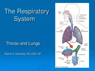
The Respiratory System Thorax and Lungs Rachel S. Natividad, RN, MSN, NP
Inspection… …Always first!!! The moment you see the patient. What position is most comfortable for him? Does he appear relaxed, anxious, uncomfortable? Is he having any trouble breathing? Assessment begins…. Tripod Position
Focused Assessment • Inspection-cont. • Color, Size and shape & symmetry of chest, any lesions or scars • Anterior Lateral Posterior
Due to deformities of the spine: Scoliosis Assymetrical chest expansion Fractured ribs flail chest Pneumothorax atelectasis paralysis of the diaphragm Asymmetrical chest Scoliosis
Increased AP:Transverse Diameter Altered size/shape:Barrel Chest
Intercostal Spaces and Muscles Retractions – indicates respiratory distress
Focused Assessment Cont… • Resp. rate (per min.) and depth (shallow, even, deep) • Normal pattern of respiration – regular rhythm • Abnormal patterns • Hyperventilation • Tachypnea vs. bradypnea • Stertorous (Noisy) • Cheyne-Stokes • Kussmaul’s • Skin: cyanosis, pallor • Nails: Clubbing • Spongy nail matrix and nail angle of greater than 160 degrees
Check for tenderness (normally nontender) Crepitus – SQ air pockets indicates air is leaking from the airways or lungs Tactile fremitus – a palpable vibration that is caused by the trasmission of air through the bronchopulmunary system. increased with fluid accumulation Abnormal if tumor, fractured ribs, chest tubes, wound site, fluid Focused Assessment Cont.:Palpation
Continuous sounds Wheezes Sibilant Sonorous (Rhonchi) Stridor Discontinuous sounds Crackles (Rales) Fine Course *Atelectic crackles Pleural friction rub Adventitious/AbnormalBreath Sounds (T 11-2) p.132
Wheezes (Continuous) Sibililant wheeze Heard 1st in expiration • high-pitched musical sounds • Due to partial blockage in airflow • Asthma, COPD, or foreign body obstruction.
Wheezes (Continuous) Sonorous wheeze (rhonchi) Heard primarily in expiration • low pitched – snoring, rattling sound • Due to air passing through large airways filled with fluid/ secretions _________________________ Stridor- partial airway obtruction viral croup, laryngeal or tracheal obstruction, epiglotottis
Interpreting what you hear… • Is the sound is continuous or discontinuous? • Is the sound occur during inhalation or exhalation, or both?
Crackles (Discontinuous) • FINE vs. COURSE Crackles • Caused by collapsed or fluid-filled alveoli popping open • usually heard in the lung bases during inhalation • Atelectic crackles • common in elderly, disappears after several deep breaths • ___________________________ • Pleural friction rub – grating sound from fluid in the pericardial space due to inflamed pleura (Pericarditis)
Abnormal Breath Sounds • Diminished breathsounds • Obese, muscular chest wall • poor inspiratory effort (elderly) • pleural effusion • Absent breath sounds • Missing lung/lobe • airway obstruction, pneumothorax- collapsed lung
Assessment Guide: Gas Exchange • Respiratory • Rate: 18 resp/min • Depth: deep, even, shallow • Effort: labored, unlabored • Breath Sounds • Describe: clear, rhonchi, inspiratory/expiratory wheezes, crackles • Location: all lobes, throughout lung fields, LLL, RUL/RML, lower lobes bilat. • Cough: present/not present • Describe: productive, moist, nonproductive • Sputum: large amount, thick yellow; moderate pink frothy sputum, sml. Amt. thin clear sputum.
Interventions in use: • Position, Turn, Cough, Deep breathe • O2 Method: nc, venti mask, rebreathing mask • Flow rate: 2L/min; 3l/min • Humidity: yes/no • Pulse Oximeter: continuous, spot monitoring • Incentive Spirometer: in use, n/a • Time used: 10 am, 11 am, 1 pm, 3 pm • Volume: 500 cc, 500 cc, 600 cc, 800 cc • Oropharyngeal Suctioning: Describe- moderate amount thick tan secretions • Med List: Albuterol inhaler, Prednisone, Theophylline


