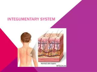
Comprehensive Study of Integumentary System Functions and Structures
480 likes | 501 Vues
This educational chapter delves into the analysis of the interdependence among integumentary, skeletal, and muscular systems in the human body pertaining to protection, support, and movement. Students will explore the structure and function of the integumentary system, covering the anatomy and physiology of the skin, different layers of the skin, functions of the integumentary system, accessory organs, healing of wounds, skin color factors, and more. Dermatologists, skin disorders, and treatments will also be discussed.

Comprehensive Study of Integumentary System Functions and Structures
E N D
Presentation Transcript
SAP2. Students will analyze the interdependence of the integumentary, skeletal, and muscular systems as these relate to the protection, support and movement of the human body. • a. Relate the structure of the integumentary system to its functional role in protecting • the body and maintaining homeostasis. Describe the Anatomy and Physiology of the Skin.
CHAPTER 6 – THE INTEGUMENTARY SYSTEM -Dermatologist: Doctor who studies and treats skin disorders I. Functions of the INTEGUMENTARY SYSTEM: A. Regulation of Body Temperature – Sweat and Blood Flow 1. Too hot: Eccrine sweat glands are activated and blood vessels dilate 2. Too cold: Arrector pili muscles contract (goosebumps) and shivering – Muscle contraction generates heat B. Protection – 1st line of defense C. Sensation – Temp., touch, pressure, pain D. Excretion – Sweat (contains urea) E. Immunity – Langerhans cells F. Synthesis of vitamin D – Aids Ca2+ and Phosphorous absorption II. STRUCTURES OF THE INTEGUMENTARY SYSTEM.
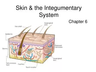
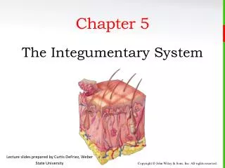
A. SKIN – Largest Organ, 22 square feet; Divided into 3 layers: 1. Epidermis –Keratinized stratified squamous epithelium a. Cell Types i. Keratinocytes (90%) - Keratinization takes 2-4 weeks ii. Melanocytes (8%) produce melanin (color and protection from UV radiation) iii. Langerhans Cells – Immune responses iv. Merkel Cells – Help sensation of touch .
b) 5 Layers of the epidermis i. Stratum Basale – Cell Division ii. Stratum Spinosum – 8-10 layers of spinelike projections iii. Stratum Granulosum: Water-Repellant sealant; Made of Lipids iv. Stratum Lucidum: Only in Palms and Soles of foot v. Stratum Corneum – Outer most layer (dead keratinocytes)
2. Dermis – “True Skin”; Contains: a. Collagen and elastic fibers i. Provides strength and elasticity ii. Striae (stretch marks) can develop when overstretching - leads to breaking of fibers b. Connective tissue, adipose, hair follicles, oil glands, and sweat glands c. Dermal Papillae – Fingerprints; For grip d. Meissner Corpuscles and other nerve endings for pain, tickling, itching 3. Subcutaneous Layer (hypodermis) – Connects Dermis to bone and muscle; Composed of Adipose Tissue
B. Accessory Organs of Integumentary System 1. Hair (Pili) – Warmth and Protection; Grows 3 years, rests 1-2 yrs a. Develops from hair follicle (group of live epidermal cells) i. Epidermal cells grow, divide, become keratinized, and die as they are moved upwards away from nutrients. ii. Hair shaft: dead cells above skin iii. Hair root: dead cells below skin iv. Arrector pili muscles: Cause “goosebumps”; Raise hairshaft (contraction generates warmth) b. Hair color determined by genes; Determines type and amount of melanin c.Disorders: i. Male pattern Baldness – Genes and testosterone; Individuals with hair follicles sensitive to DHT (dihydrotestosterone) will lose their hair in the typical balding pattern. ii.Alopecia – baldness due to autoimmune disease (immune system attacks hair follicles). Can lead to complete hair loss (eyebrows, eyelashes…) -Minoxidil (Rogaine): Vasodilator that nourishes hair follicles Propecia: Oral medication that blocks affects of DHT
9 WEEK TEST MAKEUPS?? SAP2. Students will analyze the interdependence of the integumentary, skeletal, and muscular systems as these relate to the protection, support and movement of the human body. • a. Relate the structure of the integumentary system to its functional role in protecting • the body and maintaining homeostasis. • EQ: What is the role of the accessory organs of the integumentary system?
2. Glands: a. Sebaceous Glands – i. Produces sebum – Waterproofs, keeps hair soft and pliable ii. Acne - Overactive glands become plugged and bacteria grow b. Sweat glands i. Eccrine: Produce sweat; Most prominent on palms of hands and soles of feet ii. Apocrine: In axillary region (under arm) and in groin; Become active at puberty iii. Ceruminous: Secrete ear wax iv. Mammary: Secrete milk 3. Nails – Protective coverings that contain: a. Nail plate – on top b. Nail bed (contains dividing cells) c. Lunula: Moon shaped Region over most actively growing region
III. Skin Color: Due to multiple Factors • Melanin – Protect us from UV radiation; 1. All races have same number of melanocytes; 2. Freckles = melanin patches 3. Albinism – Absence of melanin B. Carotene – Precursor of vitamin A (orangish pigment) C. Hemoglobin – Red pigment 1. Cyanosis – Shows a lack of oxygen 2. Jaundice – Buildup of bilirubin 3. Erythemia– Lots of Blood in capillaries
IV. Healing of wounds • A. Inflammation: Swelling and redness due to increased blood flow; Fluids leak into damaged tissue causing swelling • B. Shallow injuries (epidermis): Epithelial cells divide more rapidly to fill gap • C. Deep injuries (dermis or below): • 1. Blood vessels break and blood forms clot forming a scab • 2. New fibers are added by fibroblasts to bind wound together • 3. Blood vessels grow under scab, and white blood cells remove damaged tissue
V. Aging • Collagen and elastic fibers decrease, stiffen, and break • Sebaceous glands slow down (dry skin) • Sweat glands decrease production (dangers in overheating) • Number of functioning melanocytes decrease gray hair and liver spots • Skin becomes thinner and bruises more easily • Nails and hair become more brittle
VI. Skin Disorders A. Skin Cancer 1. Basal cell carcinomas – Most common; Don’t spread 2. Squamous cell carcinomas – Spread, but usually slowly 3. Melanomas – Most serious and readily spread
B. Burns 1. First degree epidermis – pain and redness 2. Second degree – red, blisters, swelling 3. Third degree – Deep tissue destruction
C. Bed sores (Decubitus ulcers) – Caused by pressure D. Contact Dermatitis – Inflammation of the skin (i.e. poison ivy) E. Corn – Inward thickening of corneum stratum F. Moles (Nevus) – Pigmented skin G. Impetigo – Contagious bacterial infection H. Warts – Viral epidermal growth I. Eczema: Scaly, oozing lesions J. Dandruff – Shedding of stratum corneum K. Hives – Itchy red patches L. Vitiligo – Loss of pigmentation M. Psoriasis – Red patches with white scales N. Hemangioma – Tumor of the skin and subcutaneous layer including blood vessels O. Acne - Blocked sebum gland P. Baldness – Combination of genetics and testosterone Q. Alopecia – Complete baldness R. Albinism – Genetic condition causing no melanin production
Contact Dermatitis HIVES
Eczema Psoriasis
Cold sore – Caused by Herpes simplex virus 1 Shingles (Caused by reemergence of Herpes Simplex virus 7 (chickenpox)
Wart Nevus (Mole) Hemangioma
Athlete’s Foot Ringworm
Pityriasis rosea • Cause is most likely viral • Not contagious • Produces a painless rash; Itching may result • Lasts 4-8 weeks; Treatment with antihistmines for itching