Download
1 / 35
500 likes | 1.3k Vues
Skin & the Integumentary System. Chapter 6. Lesson Objective. Students will know the parts of the integumentary system. Skin Hair Nails cutaneous glands. 6.1 Introduction. The cutaneous membrane commonly called skin together with certain accessory organs make up the integumentary system.

E N D
Skin & the Integumentary System Chapter 6
Lesson Objective • Students will know the parts of the integumentary system. • Skin • Hair • Nails • cutaneous glands
6.1 Introduction • The cutaneous membrane commonly called skin together with certain accessory organs make up the integumentary system. • The integumentary system consists of the skin, hair, nails, and cutaneous glands.
The skin provides a barrier against microorganisms, water, solar radiation, and others. • Upon exposure to UV radiation, the skin synthesizes an inactive form of vitamin D. • The skin is the most extensive sense organ and is important in thermoregulation.
Lesson Objective • Students will know the 4 type of membranes. • Cutaneous membranes. • Serous membranes. • Mucous membranes. • Synovial membranes.
6.2 Types of membranes The four types of membranes are: 1) Cutaneous membranes. The cutaneous membrane is the skin. This membrane is composed of a layer of epithelium over a layer of connective tissue. 2) Serous membranes. Serous membranes line body cavities (peritoneum) and surround organs such as the lung (pleura) and heart (pericardium). Do not open to the outside 3) Mucous membranes. Mucous membranes are tissues that line body cavities or canals such as the throat, nose, mouth, urethra, rectum, and vagina. Open to the outside. 4) Synovial membranes. Synovial membrane is a layer of connective tissue that lines the cavities of joints, tendon sheaths, and bursae (fluid sacs) and makes synovial fluid, which has a lubricating function.
Lesson Objective • Students will understand the basic structure of skin. • Epidermis-protection • Dermis-nourishment • Hypodermis-insulation
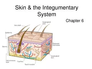
6.3 Skin & its tissues The skin, or integument, is the largest of the body organs. It includes two distinct tissue layers—the superficial layer, called the epidermis, and a deeper layer, called the dermis. A third layer, the hypodermis (sometimes called subcutaneous tissue), is not part of the skin, but serves to anchor the skin to underlying bone and muscle tissue.
Epidermis • The epidermis consists of five distinct layers, none of which contain any blood vessels. • The deepest layer of the epidermis, the stratum basale, contains cells undergoing mitosis. • The outermost layer, the stratum corneum, is composed of many layers of dead, flattened epidermal cells. • Main Function of Epidermis is PROTECTION.
Epidermis • Most of the cells in the epidermis are keratinocytes, which produce a protein mixture called keratin. • Keratinocytes are responsible for the structural strength and permeability characteristics of the epidermis. • Keratinization is the hardening of older cells. As a result, many layers of tough, tightly packed deal cells accumulate in the outer epidermis. Dead cells are eventually shed.
Other cells of the epidermis include melanocytes, Merkel cells, and Langerhans cells. • Melanocytes produce melanin, a pigment that results in dark skin color and protects the skin from ultraviolet radiation. All people have about the same number of melanocytes in their skin. Differences in the amount of melanin determine skin color. • Merkel cells are touch receptors. • Langerhans cells are macrophage. The epidermis protects underlying tissue against water loss, mechanical injury, and the effects of harmful chemicals. • A tan indicates sun damage to the skin.
Dermis • The dermis binds the epidermis to underlying tissues. The dermis consists of areolar tissue and dense irregular connective tissue. Dermal blood vessels supply nutrients to all skin cells and help regulate body temperature. • Sensory nerve fibers, hair follicles, sebaceous glands, sweat glands, and nail roots are all found within the dermis. • Main function of dermis is to NOURISH epidermis
Dermis • The strength of the dermis in mainly due to the presence of collagen fibers. • Projections of the dermis (dermal papillae) passing into spaces of the epidermis cause uneven undulations of the skin called finger prints. Genes cause fingerprints but they can change slightly as a fetus moves and presses the forming ridges against the uterine wall. This is why identical twins fingerprints are not exactly alike.
Subcutaneous • The hypodermis (subcutaneous layer) is deep to the dermis and consists of loose connective tissue and adipose tissue. Approximately half of the body's stored fat is found in the hypodermis, although the amount and location vary with age, sex, and diet. • The fat in the hypodermis functions as padding and INSULATION.
6.4 Accessory Organs of the Skin • Hair follicles • Sebaceous glands • Nails • Sweat glands • The average square inch of skin holds 650 sweat glands, 20 blood vessels, 60,000 melanocytes, and more than a 1,000 nerve endings.
Hair Follicles • Hair is present on all skin surfaces except the palms, soles, lips, nipples, and parts of the external reproductive organs. • Hair develops from a tube-like depression called a hair follicle. • The follicle extends from the surface into the dermis and contains the hair root. • As the epidermal cells divide and grow, older cells are pushed toward the surface. A hair is composed of dead epidermal cells.
A bundle of smooth muscle cells form the arrector pili muscle. Remember smooth muscle is involuntary. • If a person is emotionally upset or very cold, nerve impulses ,stimulate the muscle to contract, causing goose bumps.
Sebaceous Glands • Contain groups of specialized epithelial cells and are usually associated with hair follicles. • Holocrine glands secrete oily mixture of fatty material and cellular debris called sebum. • Sebum helps keep the hair and skin soft, pliable, and waterproof.
Many teens are familiar with a disorder of the sebaceous glands called acne. Overactive and inflamed glands in some body regions become plugged and surrounded by red elevations (pimples).
Nails • Nails are protective coverings on the ends of fingers and toes. • Each nail consists of a nail plate that overlies a surface of skin called the nail bed. • The whitish, thickened, half-moon shaped region called the lunula at the base of the nail plate is the most active growing region. • Nails grow from epithelial cells that divide and become keratinized as the rest of the nail.
Thumb nails grow the slowest, the middle nails grow the fastest.
Sweat Glands • Sweat glands are exocrine glands that are widespread in the skin. • The most numerous sweat glands, the eccrine glands, respond throughout life to body temperature elevated by environmental heat or physical exercise. Glands are common on the forehead, neck, and back. • The fluid, sweat is mostly water, but also contain salt, urea and uric acid.
Other sweat glands, called apocrine glands become active when a person is emotionally upset, frightened, or in pain. • Other sweat glands are structurally and functionally modified to secrete specific fluids such as the glands of the external ear canal that secrete earwax. The female mammary glands that secret milk are another example of modified sweat glands.
6.5 Regulation of Body Temperature • Regulation of body temp is vitally important because even slight shifts can disrupt rates of metabolic reactions. • The skin plays a key role in the homeostatic mechanism that regulates body temp. • Heat is a product of cellular metabolism. • 80% of the body’s heat escapes through the head.
Homeostasis • During intense heat (physical activity) active muscle release heat, which the blood carries away. The warmed blood reaches the brain that controls the body’s set point which signal dermal blood vessels to relax and widen (Vasodilation),heat in the blood to skin surface.
In very cold environments, the brain triggers the blood vessels to contract (vasoconstriction). If body temp continues to drop, the nervous system stimulates muscle fibers to in the skeletal muscles to contract slightly to produce heat. The body inactivates sweat glands to reduce heat loss by evaporation. If none of these raise body temp then small groups of muscles contract rhythmically with still greater force, and causes shivering, generating more heat.
6.6 Healing of Wounds • Blood vessels in affected tissues dilate and become more permeable, forcing fluids to leave the blood vessels and enter the damaged tissue. • Inflamed skin may become reddened, warm, swollen, and painful to touch. However, the dilated blood vessels provide the tissue with more nutrients and oxygen, which aids healing.
If the break in the skin is shallow, epithelial cells along the cut are stimulated to divide more rapidly, and newly formed cells fill the gap. • If the injury extends into the dermis or subcutaneous layer, blood vessels break, and the escaping blood forms a clot. The blood clot and dried tissues form a scab that covers and protects the tissue. • If the wound is extensive, the newly formed connective tissue may appear on the surface as a scar.
Common Skin Disorders • Acne- disease of the sebaceous glands that produce blackheads and pimples. • Alopecia- Hair loss, usually sudden. • Athlete’s foot- fungus infection usually in the skin of the toes and soles. • Birthmark- congenital blemish or spot on the skin, visible at birth or soon after. • Boil- bacterial infection of the skin, produced when bacterial enter a hair follicle.
Skin Disorders • Eczema- noncontagious skin rash that produces itching, blistering, and scaling. • Herpes- infectious disease of the skin, usually caused by herpes simplex virus and characterized by recurring formations of small clusters of vesicles. • Mole- fleshy skin tumor that is usually pigmented. • Psoriasis- Chronic skin disease characterized by red patches covered with silvery scales.
Skin Disorders • Scabies-disease resulting from an infestation of mites. • Ulcer- open sore. • Wart- flesh-colored, raised area caused by a viral infection.
Work Cited • Skin cross section. Image. July 8, 2012 Human+Skin.jpg khilafatworld.com • Hair Follicle picture. December 28, 2012. http://www.infovisual.info/03/037_en.html • Sebaceous gland picture. December 28, 2012. http://www.indianwomenshealth.com