Download

1 / 92
930 likes | 1.08k Vues
The integumentary system is crucial for protecting the body, serving as a barrier against environmental damage and pathogens. This system encompasses the skin, hair, nails, and glands, performing vital functions including temperature regulation, fluid balance, and sensory input. This asset explores skin anatomy, layers, various skin conditions such as burns, eczema, and skin cancer, and the healing process. It aims to foster a deeper understanding of this vital organ system for improved health awareness.

E N D
8 The Integumentary System: The Protective Covering
Multimedia Asset Directory Slide 26 Wound Repair Animation Slide 38 Degrees of Burn Animation Slide 39 Chemical Burns Animation Slide 69 Pressure Sores Animation Slide 70 Eczema Video Slide 71 Skin Cancer Video Slide 72 Decubitus Ulcers Video Slide 73 Emergency Medical Technicians Video Slide 74 Nursing Video
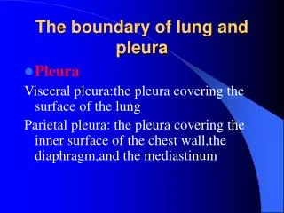
Introduction • The integumentary system protects the body from environmental damage. • The skin forms a protective barrier shielding the body from the elements and pathogens, as well as performing several other vital functions. • Skin is essential to well-being, helps to regulate body temperature, and contains many accessory organs such as nails, hair, and glands.
Learning Objectives • Discuss the functions of the integumentary system. • List and describe the layers of the skin. • Explain the healing process of skin. • Describe the structure and growth of hair and nails. • Explain how the body regulates temperature through the integumentary system.
apocrine (APP oh krine) carotene (CARE oh teen) corium (CORE ee um) eccrine (EKK rin) epidermis (ep ih DER miss) epithelial cells (ep ih THEE lee al) keratin (KAIR eh tin) keratinization (KAIR eh tin eye ZAY shun) lesion (LEE zhun) Pronunciation Guide Click on the megaphone icon before each item to hear the pronunciation.
lunula (LOO nyoo lah) melanin (MELL an in) melanocytes (MELL an oh sights) pustule (PUS tyool) sebaceous gland (see BAY shus) sebum (SEE bum) squamous cells (SKWAY mus) stratum corneum (STRAY tum core NEE um) subcutaneous fascia (sub cue TAY nee us FASH ee uh) Pronunciation Guide Click on the megaphone icon before each item to hear the pronunciation.
System Overview • The integumentary system is comprised of the skin and its accessory components including hair, nails, and associated glands.
System Overview • The integumentary system performs several vital functions. • Protection from pathogens • Balances fluid levels • Stores fatty tissue for energy supply • Produces vitamin D (with help from the sun) • Provides sensory input • Helps to regulate body temperature
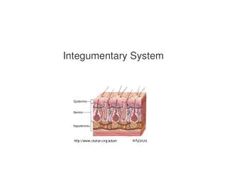
The Skin • The skin is the largest organ, weighing approximately 20 pounds and covering an area about 20.83 square feet on an adult. • A cross section of skin reveals three layers. • Epidermis • Dermis • Subcutaneous fascia
Epidermis • The epidermis is the layer of skin that we see on the outside. It is made up of five even smaller layers of tissue. • There are no blood vessels in this layer. • The cells on the surface of the epidermis are constantly shedding, being replaced with new cells that grow and arise from the deeper region called the stratum basale every 2–4 weeks.
Epidermis • The outermost layer is a layer of dead cells, called the stratum corneum, which are flat, scaly, keratinized epithelial cells. • You slough off 500 million cells every day, or about 1½ pounds of dead skin a year, allowing for rapid repair in case of injuries.
Skin Color • Specialized cells called melanocytes are located deep in the epidermis and are responsible for skin color. • Melanocytes produce melanin, a substance that causes skin color. • Variation in skin color is the result of the amount of melanin produced and how it is distributed, not the number of melanocytes. • Carotene, another form of pigment, gives a yellowish hue to skin while a pinkish hue is derived from the hemoglobin in the blood.
Effects of Disease on Skin Color • Color of skin can indicate disease. • When liver disease occurs, the body can’t break down bilirubin. The buildup of bilirubin gives the skin a deeper, yellow color. • A malfunctioning adrenal gland can cause the skin to turn bronze due to excessive melanin.
Effects of Disease on Skin Color • Excessive bruising could indicate skin, blood, or circulatory problems. • Cyanosis, or a blue coloring, results from a drop in oxygenation.
From the Streets:Skin Color • Normal skin color in light-skinned people is pink. • In dark-skinned people, inspect the mucous membranes (such as lips) to detect skin color changes. • Paleness indicates decreased blood flow through skin and can result from anemia, hypothermia or hypovolemia.
Dermis • The layer below, or inferior, to the epidermis is the thicker dermis layer. • This layer contains the following: • Capillaries • Collagenous/elastic fibers • Involuntary muscles • Nerve endings • Lymph vessels • Hair follicles • Sudoriferous glands (sweat) • Sebaceous glands (oil)
Dermis • Small “fingers” of tissue project from the surface of the dermis and anchor this layer to the epidermal layer. • Nerve fibers allow you to sense what is happening in your environment. • Vasodilation of capillaries in this layer causes blushing.
Dermis • Collagen and elastic fibers allow for the elasticity of skin, preventing the tearing of skin with movement. They allow skin to return to normal shape during periods of rest. Older people lose some of this elasticity, leading to wrinkles.
Sudiferous Glands • Two main types of sudiferous, or sweat, glands • Apocrine sweat glands secrete at the hair follicles in the groin and anal region as well as the armpits and become active around puberty and are believed to act as sexual attractants. • Eccrine glands are found in greater numbers on your palms, feet, forehead, and upper lip and are important in the regulation of temperature.
Sudiferous Glands • The body has three million sweat glands. • Sweat has no odor, but bacteria degrades the substances in the sweat over time into chemicals that give off strong smells commonly known as body odors.
Sebaceous Glands • Sebaceous glands play an important role by secreting oil, or sebum. • Sebum keeps the skin from drying out and (due to its acidic nature) helps destroy some pathogens on the skin’s surface.
Subcutaneous Fascia • The innermost layer of the skin is the subcutaneous fascia, or hypodermis. • The subcutaneous fascia is composed of elastic and fibrous connective tissue and fatty tissue. • Lipocytes, or fat cells, produce the fat needed to provide padding to protect the deeper tissues of the body and act as insulation for temperature regulation. • Fascia attaches to the muscles of the body.
How Skin Heals • If skin is punctured and the wound damages blood vessels, the wound fills with blood. Blood contains substances that cause clotting. The top part of the clot exposed to air hardens to form a scab, nature’s bandage, forming a barrier and preventing pathogens from entering. • In minor wounds, the dermis will eventually regenerate. In severe wounds, the dermis will be replaced by a scar.
Click here to view an animation on the topic of Wound Repair. Back to Directory
Burns to the Skin • Burns can be caused by heat, chemicals, electricity, or radiation. • Two factors affect assessments of damage: • Depth • Amount of area damaged
First Degree Burns • The depth of a burn relates to the layer or layers of skin affected by the burn. • First degree burns damage only the outer layer, or epidermis. • Symptoms include redness and pain, but no blister. • Pain subsides in 2-3 days and there is no scarring. • Complete healing takes about one week.
Second Degree Burns • Second degree burns involve the entire depth of the epidermis and a portion of the dermis. • Symptoms include redness, pain, and blistering. • The extent of blistering is dependent on the depth of the burn.
Second Degree Burns • Blistering extends after the initial burn. • Blisters heal within 10-14 days if there are no complications, with deeper second degree burns taking 1-3½ months. • Scarring in second degree burns is common.
Third Degree Burns • Third degree burns affect all three layers of the skin. • The surface of the burn has a leathery feel and will range in color from black, brown, tan, red, or white. • The victim feels no pain because the pain receptors are destroyed. • Also destroyed are sweat and sebaceous glands, hair follicles, and blood vessels.
Fourth Degree Burns • Fourth degree burns are the worst burns. • These burns penetrate the bone and cause bone damage.
Amount of Area Damaged • The rule of nines is used to estimate the extent of area damaged by burns. • The body is divided into the following regions, each given a percentage of body surface area value: • Head and neck – 9% • Each upper limb – 9% (2 × 9 = 18%) • Front of trunk – 18% • Back of trunk and buttocks – 18%
Amount of Area Damaged • The body is divided into the following regions, each given a percentage of body surface area value: • Front of legs – 18% • Back of legs – 18% • Perineum (including anus and urogenital region) – 1%
Figure 8-3 (continued) Assessing the degree of the burn. Bottom photos showing first degree burn (sun burn) and third degree burn.
Click here to view an animation on the topic of Degrees of Burn. Back to Directory
Click here to view an animation on the topic of Chemical Burns. Back to Directory
Burns – Clinical Concerns • The clinical concerns for burn victims relate to the functions of the skin already discussed, including: • Bacterial infections • Fluid loss • Heat loss
Burn Treatment • Severe burns require healing steps at an intensity level the body can’t manage on its own. • Damaged skin must be removed as soon as possible and skin grafting must be started. • Autografting is using the patient’s own skin, while heterografting (from a donor) is required if the patient suffered a large area of burn and has little healthy skin to graft.
Burn Treatment • Grafting requires many trips to the OR because large areas can’t be done all at once and often the grafts don’t “take.” • It is possible to grow sheets of skin tissue in the laboratory from patient cells or utilization of synthetic materials.
Nails • Specialized epithelial cells originating from the nail root form your nails. • As these cells grow out and over the nail bed, they become keratinized forming a substance similar to the horns on a bull. • The cuticle is a fold of tissue that covers the nail root.
Nails • The portion that we see is called the nail body. • Nails normally grow 1 mm every week. • The pink color of the nail comes from the vascularization of the tissue under the nails, while the white half-moon shaped area, or lunula is a result of the thicker layer of cells at the base.
Figure 8-5 Clinician performing capillary refill assessment.
Hair • Body hair is normal and serves important purposes. • Hair helps to regulate body temperature and functions as a sensor to help detect things on your skin such as bugs or cobwebs. • Eyelashes help to protect our eyes from foreign objects while hair in the nose helps filter out particulate matter.
Hair Anatomy • Visible hair is composed of fibrous protein called keratin. • The hair you see is called the shaft with the root extending down into the dermis to the follicle. • The follicle is formed by epithelial cells with a rich source of blood provided by the dermal blood vessels. • Cells divide and grow in the base of the follicle, older cells are pushed away and die, so the shaft of the hair is comprised of dead cells.
Hair Anatomy • Shaving or cutting hair doesn’t make it grow quicker or thicker. • There is a sebaceous gland associated with each hair follicle, secreting sebum that coats the hair follicle and works its way to the skin’s surface to prevent drying of the hair, acting as an antibacterial, and lubricating the hair shaft. • Sebum production decreases with age, explaining why older people have drier skin and more brittle hair.
Hair Color and Texture • Your hair color is dependent on the amount and type of melanin you produce. • The more melanin, the darker your hair. • White hair occurs in the absence of melanin. • Red hair is the result of hair that has melanin with iron in it.