
clarian/adam
Integumentary System. http://www.clarian.org/adam. Much of the text material is from, “Principles of Anatomy and Physiology, 12th edition” by Gerald J. Tortora and Bryan Derrickson (2009). I don’t claim authorship. Other sources are noted when they are used.

clarian/adam
E N D
Presentation Transcript
Integumentary System http://www.clarian.org/adam
Much of the text material is from, “Principles of Anatomy and Physiology, 12th edition” by Gerald J. Tortora and Bryan Derrickson (2009). I don’t claim authorship. Other sources are noted when they are used. Mapping of the lecture slides to the 13th edition is provided in the supplement.
Outline • Introduction • Basic structure of the skin • Accessory structures • Functions • Healing of skin wounds • Aging
Overview • Skin, hair, oil and sweat glands, nails, and somatic sensory recep-tors make-up the integumentary system. • The system helps maintain constant body temperature, provides a barrier to microbes, and provides sensory input about the external environment. • It also can mitigate, to an extent, some sources of trauma including sunlight and pollutants in the environment • The skin can serve as a window into emotions (such as in frowning and blushing). Chapter 5, page 147
Overview (continued) • Changes in skin color can indicate some types of homeostatic imbal-ances of the body. • Bluish color can indicate hypoxia, which may be associated with heart failure and other disorders. • Skin eruptions—such as chicken pox, cold sores, and measles—can reveal systemic infections and diseases involving the internal organs. • Other conditions—including pimples, moles, warts, and age spots— involve only the skin. Hypoxia = a below-normal supply of oxygen to a body tissue. Systemic = pertaining to or affecting the body as a whole. Chapter 5, page 147
Chicken Pox and Measles Measles http://123shadow.files.wordpress.com Chicken Pox http://phil.cdc.gov
Overview (continued) • Healthy-appearing skin can be important to a person’s positive self-image. • In the U.S., much time and money is spent on trying to restore skin to a youthful appearance. • Dermatology is a medical specialty involving diagnosis and treatment of disorders of the integumentary system Chapter 5, page 147
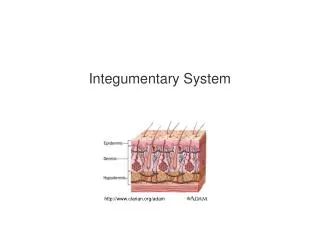
Skin • The skin—also known as the cutaneous membrane or integument— covers the external surfaces of the body. • The skin is the largest organ of the human body as measured by its total weight and surface area. Figure 5.1 Chapter 5, page 148
Skin (continued) • In adults, the skin covers an area of about 2 meters2, and weighs about 4.5-to-5 kilograms. • It ranges in thickness from 0.5 mm on the eyelids, to 4 mm on the heels of the feet. • The skin is typically about 1-to-2 mm thick on most of the body’s external surfaces. Figure 5.1 Chapter 5, page 148
Skin Structure • The skin consists of the: • Epidermis, the superficial, thinner layer of epithelial cells. • Dermis, a deeper and thicker layer of connective tissue. • Subcutaneous layer, the deepest layer made-up of areolar and adipose tissues. Figure 5.1 Chapter 5, page 148
Skin Structure (continued) http://www.cancerindex.org
Subcutaneous Layer • Fibers from the dermis anchor the skin to the subcutaneous layer. • The subcutaneous layer is attached to fascia, the connective tissue of muscles and bones. • The layer has substantial amounts of adipose tissue that give skin its form. Figure 5.1 Chapter 5, page 148
Subcutaneous Layer (continued) • The subcutaneous layer has large blood vessels that supply oxygen and nutrition to the dermis. • The layer, and to a lesser extent the dermis, have free nerve endings and Pacinian corpuscles that are sensitive to the mechanical pressure of touch. Figure 5.1 Chapter 5, page 148
Epidermis—Keratinocytes • The epidermis has four cell types: keratinocytes, melanocytes, Langer-hans cells, and Merkel cells. • Keratinocytes—about 90 percent of all epidermal cells—are organized into four or five layers, and produce the protein, keratin. • Keratin is a fibrous protein that helps to protect the skin and underlying tissues from heat, microbes, and chemicals. Figure 5.2 Chapter 5, page 149
Epidermis—Keratinocytes (continued) • Keratinocytes produce lamellar granules, which release a water-repellant compound to minimize water entry and loss. • The cells also inhibit the entry of foreign materials through the skin such as microbes and some chemicals. Figure 5.2 Chapter 5, page 150
Epidermis—Melanocytes • Melanocytes—about 8 percent of all epidermal cells—produce the pigment, melanin. • The long, slender processes of the melanocytes extend between the keratinocytes and transfer melanin to them. • Melanin is a yellow-red or brown-black pigment that helps determine skin color and absorbs ultraviolet (UV) radiation from sunlight. Figure 5.2 Chapter 5, page 150
Epidermis—Melanocytes (continued) • Once inside the keratinocytes, the melanin granules cluster to form a protective layer covering the cell nucleus on the side facing the skin surface. • Although melanin granules protect keratinocytes from UV radiation, the melanocytes themselves are susceptible to damage from excessive UV. Figure 5.2 Chapter 5, page 150
Epidermis—Langerhans Cells • Langerhans cells—which make-up a small fraction of all epidermal cells—are formed in red bone marrow and migrate to the epidermis. • The cells are involved in immune responses to microbes—however, they are readily damaged by UV radiation. Figure 5.2 Chapter 5, page 150
Epidermis—Merkel Cells • Merkel cells are the least numerous of the four epidermal cell types. • They are located in the deepest layer of the epidermis where they contact the flattened processes of Merkel disks, a type of sensory neuron. • Merkel cells and Merkel disks mediate touch sensations, especially mechanical pressure to the skin. Figure 5.2 Chapter 5, page 150
Thin Skin • Layers of keratinocytes in different stages of development form the epidermis. • In most regions of the body, keratinocytes have four layers—stratum basale (deepest), stratum spinosum, stratum granulosum, and a thin stratum corneum (shallowest). • This arrangement is known as thin skin. • Thin skin covers much of the human body, Figure 5.3 Chapter 5, page 150
Thick Skin • Where exposure to friction is the greatest—such as in the fingertips, palms, and soles—the epidermis has an additional layer of keratino-cytes. • The five layers of keratinocytes are stratum basale (deepest), stratum spinosum, stratum granulosum, stratum lucidum, and a thick stratum corneum (shallowest). • This arrangement is known as thick skin. • Anatomical details of each stratum of thin and thick skin are provided in the textbook. Figure 5.3 Chapter 5, page 150
Stratum Basale and Epidermal Growth • Newly-formed cells in the stratum basale—the deepest layer of the epi-dermis—are slowly pushed to the surface of the skin. • As the movement progresses, the cells accumulate keratin in a process known as keratinization. • The cells then undergo apoptosis. Keratinization = the process by which keratin is deposited in cells to become horny, as in the outer layer of the epidermis, and in nails and hair. Horny = composed of, or resembling, tough fibrous material consisting chiefly of keratin. Apoptosis = pre-programmed cell death in which a cell uses specialized cellular machinery to kill itself. Chapter 5, page 152
Epidermal Growth (continued) • The keratinized cells eventually slough-off, to be replaced by younger underlying cells. • The entire process takes about four weeks in an epidermis averaging 0.1 mm in thickness. Slough-off = separate from the surrounding living tissue. Chapter 5, page 152
Life and Death in the Epidermis • Epidermal cells in the stratum basale are supplied with relatively large amounts of oxygen and nutrients due to their close proximity to the cap-illaries in the dermis. • These cells are metabolically active, and undergo continuous mitotic cell division to produce new keratinocytes. • The layers of the epidermis overlying the stratum basale receive less oxygen and nutrients, and the cells eventually die. Chapter 5, page 152
Rate of Cell Division • The rate of cell division in the stratum basale increases when the outermost layers of the epidermis are damaged such as by abra-sions or burns. • The mechanisms controlling the rate of mitotic cell division are not well-understood. • Hormone-like proteins including epidermal growth factor have roles in this process. Chapter 5, page 152
Epidermal Tidbits • Constant exposure to skin friction results in the formation of a callus, an abnormal thickening of the outermost layer of epidermis (stratum corneum). • An excessive number of keratinized cells shed from the scalp is known as dandruff. Chapter 5, page 152
Dermis • After the epidermis, the next deeper part of the skin is the dermis—it consists of connective tissue composed of collagen and elastic fibers. • The network of fibers has substantial tensile strength, and the ability to stretch and recoil easily. • The dermis contains a relatively small number of fibroblasts, macro-phages, and adipocytes near its boundary with the underlying subcu-taneous layer. Tensile strength = the amount of longitudinal mechanical force a tissue can withstand without tearing apart. Chapter 5, page 152
Dermis (continued) • Blood vessels, nerves, exocrine glands, and hair follicles are embedded in the dermis. • The dermis is essential to the epidermis, and together they have close structural and functional interrelationships. • The dermis consists of a superficial papillary region and a deeper reticu-lar region. Chapter 5, page 152
Dermis—Papillary Region • The papillary region, which makes-up about one-fifth the thickness of the dermis, consists of areolar tissue of collagen and elastic fibers. • Dermal papillae are finger-like projections into the undersurface of the epidermis. • Some of these papillae contain capillary loops that serve as a source of blood supply (and therefore oxygen and nutrients) for the epidermis. Figure 5.1 Chapter 5, page 152
Dermis—Papillary Region (continued) • Other dermal papillae contain sensory receptors including Meissner corpuscles and free nerve endings. • Meissner corpuscles are sensitive to the mechanical pressure from touch. • Free nerve endings are sensitive to hot, cold, pain, tickling, and itch-ing. Figure 5.1 Chapter 5, page 153
Dermis—Reticular Region • The reticular region, attached to the underlying subcutaneous layer, consists of connective tissue of fibroblasts, collagen, and elastic fibers. • The collagen fibers are interlaced in a net-like arrangement. • Some adipose cells, hair follicles, nerves, sebaceous glands, and sudoriferous glands are found in the space between the collagen fibers. Sebaceous gland = produces an oily secretion. Sudoriferous gland = produces perspiration and secretes it at the surface of the skin. Figure 5.1 Chapter 5, page 153
Dermis—Reticular Region (continued) • Collagen and elastic fibers in the reticular region give skin its strength, elasticity, and extensibility. • Signs of extensibility can be seen around the joints, and in pregnancy and obesity. • Extreme stretching of the skin can produce small tears in the dermis, producing striae, or stretch marks. • The striae are visible as red or silvery-white streaks on the surface of the skin. Extensibility = the capability of being stretched. Elasticity = the property of returning to an initial form or state following deformation. Figure 5.1 Chapter 5, page 153
Epidermal Ridges • The surfaces of the palms, fingers, soles, and toes have patterns that form from epidermal ridges. • They appear as straight lines or as a pattern of loops and whorls such on the fingertips. • The ridges are produced during the third month of fetal development from downward projections of the epidermis into the dermis between the dermal papillary. Figure 5.1 Chapter 5, page 153
Epidermal Ridges (continued) • Epidermal ridges increase the surface area of the epidermis, and there-fore the grip of hands and feet by increasing the amount of friction with objects and surfaces. • Imagine having entirely smooth hands when trying to hold a glass of water. Figure 5.1 Chapter 5, page 153
Fingerprints and Footprints • The ducts of sweat glands open as sweat pores onto the epidermal ridges. • The ridges and sweat form fingerprints or footprints upon contact with a smooth surface. Figure 5.1 Chapter 5, page 153
Fingerprint Card http://library.kentlaw.edu A digital (electronic) method known as LiveScan is now used.
Genetic Uniqueness • The epidermal ridge pattern is genetically-determined and unique to each individual. • The pattern usually does not change during a lifespan—except to enlarge—which makes it very useful for identification purposes. • The scientific study of epidermal ridge patterns is known as derma-toglyphics. Chapter 5, page 153
Nourishment • The dermal papillae greatly increase the area of contact between dermis and epidermis. • An extensive network of blood vessels in the dermis serves as the source of nutrition for the overlying epidermis. • Molecules can diffuse from capillaries in the dermal papillae to the cells of the basal stratum in the epidermis. • The nourishment enables epithelial stem cells to divide via mitosis, and keratinocytes to grow and develop. Chapter 5, page 153
Keratinocytes migrate toward the skin surface and away from the blood source. • They are eventually no longer able to obtain nutrition, which leads to the breakdown of their organelles. • The keratinocytes continue their migration to form the outer layer of dead cells in the epidermis. Chapter 5, page 153 Cell Migration
Epidermal Junctions • The dermal papillae closely fit with the epidermal ridges to form epider-mal junctions between the dermis and epidermis. • This structure enables the skin to resist shearing forces that could other-wise separate the epidermis from the dermis. Shearing force = a mechanical force applied perpendicular or tangential to the face of a tissue. Chapter 5, page 153
Skin Color • The three pigments that produce the full range of skin colors are melanin, hemoglobin, and carotene. • The concentration of melanin produces skin colors from pale yellow, to reddish-brown, and to black. • Melanin-producing cells, known as melanocytes, are most plentiful in the epidermis of the penis, nipples of the breast, areolae (area surrounding the nipples), face, and limbs. • Melanocytes are also found in large concentrations in the mucous mem-branes. Mucous membrane = mucus-secreting membrane lining all body cavities or passages that communicate with the exterior. Chapter 5, page 153
Skin Color Variations • The number of melanocytes is about the same in all people of all heri-tages. • The differences in skin color are primarily due to the amount of mela-nin that the melanocytes can produce and transfer to keratinocytes in the epidermis. • Darker-skinned individuals have high concentrations of melanin in the epidermis. • Lighter-skinned individuals have lower epidermal concentrations of melanin. • Skin color also has an environmental component, as discussed during the biology review. Chapter 5, page 154
A Few Skin Color Variations http://anthro.palomar.edu http:/cache.eb.com
Visual Appearance of Light Skin • The epidermis can appear translucent in individuals who have light-colored skin. • The skin color can range from pink-to-red depending on the oxygen content of the blood in the capillaries of the dermis. • This color is due to hemoglobin, the oxygen-carrying pigment in red blood cells. Translucent = semi-transparent; features on the other side can be seen, but are not distinguishable. Chapter 5, page 154
Melanin Accumulation • Melanin accumulates in patches known as freckles in some people. • Age spots, ranging in color from light brown to black, are also accumula-tions of melanin. • A round, flat, or raised area of localized growth of melanocytes is known as a nevus, or mole. Chapter 5, page 154
Freckles http://blogs.cornell.edu
Age Spots and Moles Mole http://img.webmd.com Age spots http://images.meredith.com
Moles versus Melanomas http://healthcare.utah.edu Melanomas, a type of carcinoma, are often readily-treatable if detected early. It’s good to know the warning signs.