Lumbar Sympathetic Block
A useful procedure for chronic pain


Lumbar Sympathetic Block
E N D
Presentation Transcript
Lumbar sympathetic block Dr KritikaDoshi MD, DA, FIPP, ISSP (Pain fellowship) Consultant In Chronic Pain: Doshi pain Relief Centre, THANE Bethany Hospital, Jupiter Hospital www.painreliefcentre.in
History • 20th century: John Langley coined the term- Autonomic NS to describe the unconscious function of the internal organs • Shortly later, sympathetic and parasympathetic as components maintaining homeostasis was recognized Langley JN; The autonomic nervous system. Brain 1903;26:1-26
Lumbar Sympathetic Block • Well established intervention • 75 yrs since first reported by Brunn and Mandl in 1924- selheim’s tech for paravertebral block- for visceral pain relief • Kappis reported benefit of surgical resection of lumbar symp nerves
1950- Bonica, Moore studied the benefit of LSB in Rx of causalgia and post-traumatic reflex dystropy of WWII servicemen • 1970- Reid described the lateral approach avoiding the transverse process • 1988-89 Pernak and Sri Kantha described the RF technique
Lumbar Sympathetic Block • The sympathetic nervous system has been implicated in numerous pain syndromes ranging from neuropathic pain to vascular pain to visceral pain. In some instances, its role in other painful conditions such as headaches and musculoskeletal pain has been explored. Local anesthetic blockade of sympathetic ganglia has become common practice among pain practitioners to diagnose and treat sympathetically mediated pain (SMP) When pain relief has been afforded with the initial block, chemical and thermal neurolysis has been performed in the attempt to provide prolonged analgesia.
Mechanism of Pain Relief • Symp efferents are accompanied by visceral sensory afferents- symp block interrupts nociceptive signals from viscera to brain • Ischemic pain- relief due to vasodilatation in vasospastic and vasoocclusive conditions. Smithwick RH: The rationale and technique of sympathectomy for relief of vascular spasm of the extremities. N Engl J Med 1940;222:699-703 • Eliminates nor-epinephrine mediated activation of nociceptors
ANS: central and peripheral portion • Efferent pathway: i) primary presynaptic/preganglionic neurons ii) secondary postsynaptic/postganglionic neurons Motor neurons of ANS located in periphery
Sympathetic nervous system • Cell bodies of preganglionic fibers: intermediolateral horn of SC from T1-L2 • Efferent fibres travel from SC along ventral roots • Each fibre has multiple synapses with postganglionic cells • Wrc myelinated, grc nonmyelinated
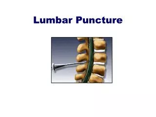
Anatomy • The lumbar sympathetic chain lies at the anterolateral border of the vertebral bodies • There is variation in the size, number, and position of the ganglia wrt the vertebral bodies • Vascular structures in close proximity: • i) The aorta is positioned anteriorly & medial to the left sympathetic chain • ii) The IVC is close to the chain on the right in an anterior plane • Iii) Many other small lumbar arteries and veins are positioned near the sympathetic chain
Applied anatomy Sympathetic chain enters the lumbar retroperitoneal space under crus of diaphragm Anteriorly: Parietal reflection of peritoneum Posteriorly: Periosteum overlying vertebral body, fibroaponeurotic origin of psoas and its fascia Laterally: genitofemoral nerve on the psoas Posterolaterally: Kidney and ureter Anteromedially: Aorta on left, IVC on right
Sympathetic Paravertebral: ganglia • segmentally arranged along anterior margin of vertebral column ( embryonically- 1 ganglion per spinal segment; many fuse) • Cervical : 3 • Thoracic: 10-12 • Lumbar : 3-5 • Sacral : 4 pairs, single Ganglion Impar
Lumbar Ganglia • 3-5 on either side (L1,L2 usually fused) • 5-15mm in size Rocco AG, Palomgi D, Raeke D: Anatomy of the lumbar sympathetic chain. Reg Anes 20:13-19,1995 • Fusiform to round in shape • L2: lower 1/3 of body • L3: upper 1/3 of body • L4: variable- usually middle of body
The ganglia on the two sides of the body are rarely symmetric in their appearance, branches, number, and location: Yeager G.H. and R.A. Cowley- Anatomical observations on the lumbar sympathetics with evaluation of sympathectomies in organic peripheral vascular disease. Ann. Surg. 127:953-967, 1948
The L-1 vertebral body usually has no ganglion. • The L-2 ganglion is furthermost back from the anterior border of the vertebral body, at a distance of 12-13 mm at the junction of the upper two thirds and the lower one-third • The L-3 ganglion is the highest where the ganglion is almost at the anterior border at the junction of the upper one-third and lower two-thirds. • The L-4 ganglion is more posterior to the anterior border and is very variable in its relationship to the discs. • The L-5 ganglion is further posterior, but again extremely variable. • If one compares 2 sides, the locations may be different. • The most predictable sites are L-2 and L-3 ganglia.
Preganglionic fibres pass as the white rami communicantes along the corresponding spinal nerves to the paravertebal ganglia • These fibers interface with postganglionic efferents and also on the opposite ganglia
Indications • Complex Regional Pain Syndrome (CRPS), types I and II Neuropathy Phantom limb pain Arterial insufficiency of the lower extremity Raynaud’s syndrome Postherpetic neuralgia Cancer pain Ischemic pain Hyperhidrosis Frostbite Lower extremity crush injury
Indications • Vascular conditions LL: arteriosclerosis, diabetic gangrene, Buerger’s disease, Raynaud’s phenomenon, reconstructive vascular Sx after arterial embolic occlusion, frostbite • SMP: CRPS I,II • Misc: hyperhydrosis, amputation stump pain, intractable urogenital pain, trench foot • Discogenic pain
Contraindications • Presence of infection, coagulopathy • Anatomic abnormality which may interfere with procedure
Site of blockade • L2: lumbar pain • L3: axial pain in spine • L4: lower limb • L4/5,: ankle / foot
Procedure technique • Prone/lateral • Classic- paravertebral / lateral approach • 1/2/3 needle technique • blind / CT-guided / Fluoroscopy guided • Diagnostic / therapeutic / neurolytic / RF • Continuous catheter in paravertebral space
Protocol for Intervention • A radiolucent operating table can facilitate and reduce the time required to perform the procedures. • For all procedures at the lumbar level it is important to decrease the lumbar lordosis, which can be done by placing a pillow under the patient’s abdomen with the patient in prone position. • AP view is obtained. After aligning the end plates of the vertebral bodies by cephalic or caudal tilt, the C-Arm of the fluoroscopic machine is rotated to the Right or Left for oblique views to obtain a “Scotty” view. The SAP, TR and the pedicle are identified. • The needle is directed to the target point under fluoroscopic control and small amount of contrast agent is injected to confirm correct needle placement and rule out vascular uptake before injecting any medication. • To perform the procedures safely a “3D” approach is essential. The three D’s stand for Direction of the needle in AP or Oblique view, Depth of the needle in Lateral view and again Direction of the needle in AP or Oblique view
Procedure From Waldman SD: Interventional Pain Management, 2nd ed. Philadelphia, WB Saunders, 2001, p 487 • Standard monitoring • IV access • Prone position, pillow under abdomen • Selected vertebral level: square vertebra • C-arm directed obliquely to get “scottie-dog”: TP flush with body of vertebral body • Entry point is just inferior to inferior border of tip of TP
Needle is advanced in gun-barrel approach till LOR is felt • Needle tip is confirmed in AP/lat view by injecting non-ionic, water soluble dye • Contrast appears along anterolateral vertebral border in a tight, linear fashion without any lateral or posterior extension
Drug • Diagnostic: LA (0.75% Ropivacaine, 0.5% Bupivacaine or 2% Lignocaine) • 1ml aliquots, preservative free drug after r/o vascular uptake under live fluro • Single needle: at L3; vol=15-20 ml • Double needle: at L2, L4; vol=10ml • 3 needle: at each level; 5-7ml
v • The Lumbar Sympathetic ganglia are located on the surface of psoas muscle
RF ablation • Procedure same • Lower 1/3 L2; upper 1/3 L3; middle L4 • 15cm, 10mm active tip RF needle • Sensory st: 50Hz; vague discomfort in back at 0.2-0.5V. Reposition if paresthesia in groin – gf nerve • Confirm absence of motor st in LL at 2Hz, upto 2V • Inject 1ml 0.75% Ropivacaine + 20mg Kenacort • Lesion at 80*C for 90sec- 2 lesions with tip facing cephalad and caudad
Neurolytic blocks • 2/3 needle technique at L2, L3 • Less volume of drug • 2-5ml adequate • 6-10% phenol preferred • 50-100% alcohol • Drug to be mixed with dye to confirm no lateral diffusion of drug (neurolysis- only after diagnostic block +ve and for long lasting relief)
Single needle (diagnostic) • Line joining costal margins bilat: L1-2 • Intercristal line: L4-5 • 4-5cm away from L2 spinous process • 15cm needle, cephalad, 45*, till TP • Needle withdrawn, redirected 85* till 4cm beyond marking till LOR • Negative aspiration for blood, CSF
The distance between the SP & TP and TP to the sympathetic ganglia is constant :4-5 cm • (the AP dimension of the lumbar vertebrae varies < 0.6 cm irrespective of the patient's build) 4-5cm 5cm
Post procedure- Adequacy of block Bed-side tests: • Increase in temperature: thermography 1*c change, ( LL temp measured at ant thigh, medial aspect of leg, dorsum of foot, great toe) • Vasodilatation – superficial skin blood flow measured by laser doppler imaging
Sweat test- ninhydrin test cobalt blue test starch iodine test • Decreased edema • Decreased pain- if SMP (Clinically pain relief outlasts duration of LA)
Sequale/ side effect • Hypotension • Back Pain • Sudomotor paralysis causing dry skin • Pathologic gustatory sweating- Mailis A, Furlan A. Sympathectomy for neuropathic pain. Cochrane database Syst Rev 2003;2:CD002918
Complications • Back pain: needle puncture through back muscles • Somatic nerve irritation/blockade- chemical neurolysis- commonly with genitofemoral or lumbar plexus if needle is too posterior or lateral (L4-5) • Renal/ureteric trauma: needle placement is too lateral • No imaging used- neuraxial injury/injectionFailure of ejaculation- bilat blockade • Intravascular injection resulting in toxic, systemic reactions • subarachnoid injection- into dural sleeve ( small aliquots injected) • Inadvertent intervertbral disc puncture
conclusion • Effective, safe method • Results comparable to chemical and surgical neuroablation. • Precise neurolysis with radiofrequency lesioning decreases the risks of sympathectomy • Radiofrequency sympathectomy also allows repeat procedures without the risk of distorting the original anatomy