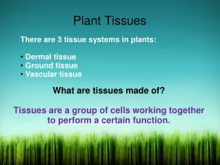
Plant Tissues
Presented by, K.Sharath Deepika B.Sc (B.Z.C) II nd year. Plant Tissues. A tissue is an ensemble of similar cells and from the same origin, that together carry out a specific function. These are called tissues because of their identical functioning.

Plant Tissues
E N D
Presentation Transcript
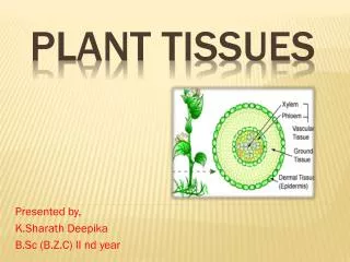
Presented by, K.Sharath Deepika B.Sc (B.Z.C) II nd year Plant Tissues
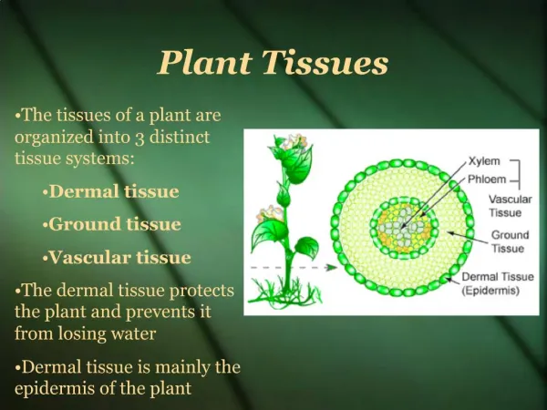
A tissue is an ensemble of similar cells and from the same origin, that together carry out a specific function. • These are called tissues because of their identical functioning. • Organs are then formed by the functional grouping together of multiple tissues. K.Sharath Deepika What is a Tissue?
K.Sharath Deepika Tissues Meristematic Tissues Permanent Tissues Shoot Apex Root Apex Simple Complex Special
Meristematic tissue is the composition of the meristematic cells. • The Meristematic cells are usually thin walled living cells with dense cytoplasm and a large nucleus. • Meristemsare called the group of young cells that have capacity to divide into new cells. K.Sharath Deepika Meristematic Tissue
Kinds of meristems: • Apical meristems –found at the tip of stems & roots - types: 1. Shoot Apex, 2.Root Apex. • Lateral meristems –cambium - found along the sides of roots & stems - increase width or diameter of stems & roots - types: 1. vascular cambium 2. cork cambium • Intercallarymeristems –found at the bases of young leaves & internodes - responsible for further lengthening of stems & leaves K.Sharath Deepika Kinds of Meristems
K.Sharath Deepika Shoot Apical Meristem
K.Sharath Deepika Root Apical Meristem
K.Sharath Deepika Lateral Meristem Stem in cross section 1,3 year old stems Basswood Root – Cross Section
K.Sharath Deepika Intercalary meristem
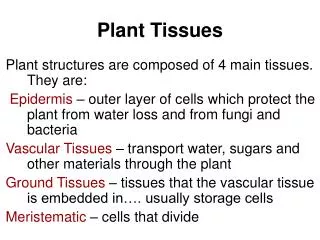
A Permanent Tissue is formed by the division and differentiation of meristematic cells. • The cells of these tissues have lost the power of division permanently. • Permanent Tissues can be classified into three types – (i) Simple, (ii) Complex, (iii) Special Tissues. K.Sharath Deepika Permanent Tissues
K.Sharath Deepika Permanent Tissues Simple tissues Complex Tissues Special Tissues Xylem Phloem Parenchyma Collenchyma Sclerenchyma Secretory cavities Laticiferous Tissues Digestive Glands NectarGlands Hydathodes Osmophors
A group of similar cells that perform a common function is called a Simple Tissue. • It is of three different types, they are - Parenchyma - Collenchyma - Sclerenchyma K.Sharath Deepika Simple Tissues
Parenchyma is the most basic type of cells. • The cells are usually isodiametric. • Parenchyma is seen in the complex tissues like Xylem and Phloem. • There are five types of parenchyma. • Aerenchyma – large intercellular spaces • Chlorenchyma –presence of chlorophyll. • Prosenchyma –elongated cell that give mechanical support • Storage Parenchyma –stores food material • Water storage Tissue –stores water (Succulents) K.Sharath Deepika Parenchyma
K.Sharath Deepika Chlorenchyma Aerenchyma Storage Parenchyma in Bean
Collenchyma is the living Tissue which gives tensile strength. • It is commonly found below the epidermis. • The cell walls are thick and lignified. • There are three types of collenchyma • Lamellar collenchyma –cell arrangement is in tangential rows • Lacunar Collenchyma –Cells have large intercellular spaces called ‘Lacuna’. • Angular Collenchyma –wall deposition is at corners of the cells. K.Sharath Deepika Collenchyma
K.Sharath Deepika collenchyma
Sclerenchyma is a dead tissue that gives mechanical support. • The cell wall is made of cellulose and lignin. • They are of two types: • Fibres –elongated cell with tappering ends. (i) Xylary fibres – present in xylem (ii) Extra xylary fibres –present other than xylem • Sclereids –short and are irregular in shapes. Ex: astro sclerieds, osteo sclereids etc. K.Sharath Deepika Sclerenchyma
K.Sharath Deepika sclerenchyma
Parenchyma plays a vital role in Photosynthesis, Respiration, Storage and Secretion. • Parenchyma gives turgidity to the young plants. • Collenchyma gives flexibility an elasticity to the plant. • Sclerenchyma protects the plant from stretching, bending, weight, pressure. K.Sharath Deepika Functions of Simple Tissues
Tissues with different kinds of cells perform similar function. • They are mainly helpful in conduction. • There are two main types: • Xylem • Phloem K.Sharath Deepika Complex Tissues
Xylem in the water conducting tissue and also provides mechanical support. • It originates from two sources like procambium and the vascular cambium. • The first formed cells are called protoxylem and the latter are called metaxylem elements. K.Sharath Deepika Xylem
It is composed of three types of cells. • Tracheary elements –There are dead cells, conduct water. (i) Tracheids: elongated with tappering ends. (ii) Vessel elements: wide and cylindrical structures. • Vessel Elements –They are wide and cylindrical. • Xylem Fibres –They are dead with thick lignified walls. • Xylem Parenchyma –normal parenchyma cells. (i) Axial Parenchyma – develops from fusiform cells (ii) Ray Parenchyma –develops from ray initials. K.Sharath Deepika Structure of Xylem
K.Sharath Deepika Xylem elements
Phloem conducts water, minerals and gives mechanical support. • It originates from two sources like procambium and vascular cambium. • The first formed cells are called protophloem and latter are metaphloem. K.Sharath Deepika Phloem
It is composed of four types of cells. • Sieve Elements – they are living with sieve areas. (i) Sieve cells – With unspecialized sieve areas. (ii) Sieve tube –With Specialized Sieve areas. • Companion cells –elongated cells which support sieve tubes. • Phloem Fibres –dead cells with tappering ends. • Phloem Parenchyma –Living and thin walled cells similar to parenchyma (i) Axial Parenchyma – develops from fusiform cells (ii) Ray Parenchyma –develops from ray initials. K.Sharath Deepika Structure of Phloem
The cells or tissues that are concerned with secretion or excretion from the plant body. • They are located in different parts of the plant body and are widely distributed. • Many plant secretions are of high considerable economic importance. • They include rubber, gums, oils, resins and mucilage. K.Sharath Deepika Secretory (or) Special Tissues
1.Digestive glands:- • They are found in insectivore plants and secrete proteolytic enzymes. Ex:- Nepenthes – digestive glands are spherical, multicellular. K.Sharath Deepika Types of Special Tissues Pitcher in Nepenthes Glandular hairs in Pinguicula
2. Nectar Glands:- • They are called as nectaries. • They secrete sugary substance called nectar which attracts insects and promote pollination. (i) Floral Nectaries – In floral regions (ii) Extra floral Nectaries –Other than floral regions. Ex:- Dianthus K.Sharath Deepika Nectar glands in Dianthus
3. Osmophors:- • They are special glands which produce volatile essential oils and impart fragrance to flowers. • The osmophors vary in structure for different species i.e. flaps, cilia, brush etc. • They promote Cross Pollination. Ex:- Orchids K.Sharath Deepika Flap like osmophors in Caryanthesmacranthes
4. Secretory Cavities:- • The secretions released are stored in the spaces within the gland. • These are formed by the breakdown of secretory cells. (i) Lysigenous cavities –formed by the death of the secretory cells. (ii) Schizogenous cavities –formed by the enlargement of intercellular space between secretory cells. Ex:- Eucalyptus. K.Sharath Deepika Oil glands in Eucalyptus leaf section
5. Hydathodes:- • Hydathodes are also called as water stomata. • They become active when root pressure increases due to reduced transpiration. • Water is forced out of tracheids in the form of drops through water pores. • This process is called guttation. Ex:- Lycopersicon K.Sharath Deepika Guttation in Lycopersicon esculentum
6.Laticiferous Tissues:- • Laticifers are specialize parenchyma cell which secrete a viscous fluid, known as latex. • Latex is mostly white in colour but sometime colour. (i) Laticiferous cells:- They are isolated, elongated, slender. (ii) Laticiferous Vessels:- They are formed by series of cells whose wall break an form canals. Ex:-Preparation of rubber from (Ficuselastica) K.Sharath Deepika Collection of latex from Ficuselasticato prapare Rubber
www.wikipedia.org – Tissue, Types of Tissue – 23/12/2012. • www.googleimages.com – tissue pictures -23/12/2012 • Common core a textbook of Botany – Special Tissues – 23/12/2012. K.Sharath Deepika Reference