Skin
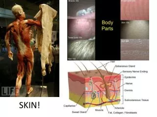
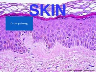
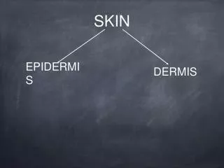
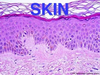
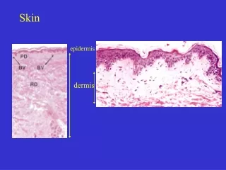
4 week. Skin It consists of epidermis which is the superficial epithelial tissue. It derived from surface ectoderm. The dermis is a deeper layer composed of dense irregularly arranged connective tissue which is derived from mesoderm.

Skin
E N D
Presentation Transcript
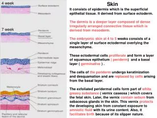
4 week Skin It consists of epidermis which is the superficial epithelial tissue. It derived from surface ectoderm. The dermis is a deeper layer composed of dense irregularly arranged connective tissue which is derived from mesoderm. The embryonic skin at 4 to 5 weeks consists of a single layer of surface ectodermal overlying the mesenchyme. These ectodermal cells proliferate and form a layer of squamous epithelium ( periderm) and a basal layer ( germinative ) . The cells of the periderm undergo keratinization and desquamation and are replaced by cells arising from the basal layer. The exfoliated peridermal cells form part of white greasy substance ( vernix caseosa ) which covers the fetal skin. Later, the vernix contain sebum from sebaceous glands in the skin. This vernix protects the developing skin from constant exposure to amniotic fluid with its urine content. Also, it facilitates birth because of its slipper nature. 7 week
11 weeks Newborn The basal layer of the epidermis becomes the stratum germinativum which produces new cells that are displaced into the layers superficial to it. By 11 weeks, cells from the stratum germinativum have formed an intermediate layer. Also, it forms epidermal ridges ( 10 week ). Replacement of peridermal cells continues until about the 21 week, after that, the periderm disappears and the stratum corneum forms.
Proliferation of stratum germinativum cells form epidermal ridges which extend into the developing dermis. This ridges begin to appear at 10 weeks and are permanently established by the 17thweek. These epidermal ridges produce grooves on the surface of the palm; sloe and digits. The pattern of development of these epidermal ridges is determined genetically and constitutes the basis for examining fingerprints in criminal investigations and medial genetics. Dermatoglyphics: It is the study of the patterns of the epidermal ridges of the skin. It is used for diagnosis of Down syndrome.
Late in the embryonic period, neural crest cells migrate into the mesenchyme of the developing dermis and differentiate into melanoblasts. Later these cells migrate into the dermoepidermal junction and differentiate into melanocytes. This differentiation involves the formation of pigment granules. The melanocytes begin producing melanin before birth. The relative content of melanin in the melanocytes accounts for different colors of skin. Skin is classified as thick or thin according to the thickness of the epidermis. Thin skin covers most of the body. It contains hair follicles; arrector pilli muscles of hairs; sebaceous glands and sweat glands. Thick skin covers the palm and soles. It has only sweat glands.
Dermis Most of the mesenchyme that differentiates into the connective tissue of the dermis originates from the somatic layer of the lateral mesoderm. Some of it is derived from the dermatomes of the somites. By 11 weeks, the mesenchymal cells have begun toproduce collagenous and elastic connective tissue fibers. The dermal ridges appear at the same time of epidermal ridges appearance ( 10 week ) and interdigitate with each other. Capillary like vessels have been observed in the dermis at the end of the 5th week. Capillary loops develop in some of these ridges. Some capillaries acquire muscular coats and become arterioles and arteries. Sensory nerve endings are formed in the other ridges. Afferent nerve fibers are responsible about papillary ridge formation. By the end of the 1st trimester, the major vascular organization of the fetal dermis is formed.
Sebaceous Glands Most sebaceous glands develop asbuds from the sides of the developing epithelial root sheaths of hair follicles. The central cells of the alveoli break down, forming an oily secretion ( sebum ). Sebaceous glands may develop as buds from the epidermis, they are called sebaceous glands independent of hair. They are present in the glans penis and labia minora.
Sweat Glands Eccrine sweat glands are located in the skin through out most of the body. They develop as epidermal downgrowths into the underlying mesenchyme. As the buds elongates, its end coils to form the primordium of the secretory part of the gland. The peripheral cells of the secretory part of the gland differentiate into myoepithelial cells and secretory cells. Myoepithelial cells are specialized smooth muscle cells. The eccrine sweat glands begin to function shortly after birth. The apocrine sweat glands develop from downgrowths of the stratum germinativum of the epidermis that give rise to hair follicles. So, their ducts open into the upper part of the hair follicles superficial to the openings of the sebaceous glands. These apocrine glands are present in the axilla, pubic and perineal regions. They secrete during puberty.
Hair It begin to develop early in the fetal period ( 9th to 12 week ). It becomes recognizable at about the 23 week. At first, it recognizes on the eyebrows; upper lip and chin by the end of the 12th week. A hair follicle begins as a proliferation of the stratum germinativum of the epidermis and extends into the underlying dermis. The hair bud becomes club- shaped, forming the hair bulb ( primordium of a hair root ). The hair bulb is invaginated by a small mesenchymal hair papilla. The epithelial cells of the hair bulb constitute the germinal matrix which laterproduces the hair. The peripheral cells of the developing hair follicle form the epithelial ( epidermal ) root sheath and the surrounding mesenchymal cells differentiate into the dermal root sheath. As cells in the germinal matrix proliferate, they are pushed toward the surface, where they become keratinizing to form the hair shaft.
Lanugo is the first hairs that appear toward the end of the 12th week and is plentiful by 17 to 20 weeks. It is fine; soft and lightly pigmented. This hair persists over most of the body except in the axillary and pubic regions where it is replaced at puberty by coarser hairs. The melanin is produced by melanocytes and is transferred to the hair- forming cells in the germinal matrix several weeks before birth. This melanin accounts for different hair colors. Arrector muscles of hairs differentiate from the mesenchyme surrounding the hair follicle and attached to the dermal root sheath of the hair follicles and the papillary layer of the dermis. These muscles are poorly developed in the hairs of axilla and in certain parts of face. The hairs of eyebrows and cilia forming eyelashes have no arrector muscles.
Nails Toenails and figernails begin to develop at the tips of the digits at about 10 weeks. Development of the fingernails precedes the toenails by about 4 weeks. The primordia of nails appear as thickened areas of epidermis at the tip of each digit. The nail fields are surrounded laterally and proximally by folds of epidermis ( nail folds ). Cells from the proximal nail fold grow over the nail field and become keratinized to form the nail plate. At first the nails are covered by eponychium ( superficial layer of epidermis ). This degenerates except at the base where cuticl is formed. The skin under the free margin of the nails is the hyponychium. The fingernails reach the fingertips by about 32 weeks. The toenails reach the toetips by about 36 weeks. Nails that have not reached the tips of the digits at birth indicate prematurity.
Mammary Glands They are modified and specialized type of sweat glands.The mammary crests appear during the 4th week. These crests are thickened strips of ectoderm, extending from the axillary to inguinal regions. It persists only in the pectoral region. Mammary buds begin to develop during 6th week as solid downgrowths of the epidermis into the underlying mesenchyme. The mesenchyme is an inductive for this. Each primary bud gives rise to several secondary buds that develop into lactiferous ducts and their branches.
Canalization of these buds is induced by placental sex hormones. By full term, 15 to 20 lactiferous ducts are formed. The fibrous connective tissue and fat develop from the surrounding mesenchyme. During late fetal period the epidermis at the site of origin of the mammary gland becomes depressed forming a shallow mammary pit. In newborn infants the nipple is depressed. After birth the nipples rise from the mammary pits due to proliferation of the surrounding connective tissue of the areola. The smooth muscle fibers of the nipple and areola differentiate from surrounding mesenchymal cells. Full development occurs at about 20 years.
The mammary glands develop similarly in both sexes. Some secretion ( witch’s milk) may be produced. These occur by maternal hormones passing through the placental membrane into fetal circulation. Only the main lactiferous ducts are formed at birth and it underdeveloped until puberty. In female, the breasts enlarge due to development of the mammary glands and the accumulation of fat associated with them. Growth of the duct system occurs because of the raised levels of circulating estrogens, prolactin, corticoids and growth hormone. If pregnancy occurs, the mammary glands complete their development. The intralobular ducts undergo rapid development forming buds that become alveoli. The breasts become hemisherical in shape and largely because of the deposition of fat.
Supernumerary Breast and Nipples An extra breast ( polymastia ) or nipple ( polythelia ) occurs in about 1 % of female population and is an inheritable condition. They usually develops just inferior to the normal breast. They less commonly appear in the axillary or abdominal regions of females. They develop from extra mammary buds that develop along the mammary crests. They become obvious in pregnant women. Polythelia are common in males, they are mastaken for moles. About one third of affected persons have 2 extra nipples or breast. Polymastia very rarely occurs in a location other than along the course of the mammary crest. It develops from tissue that was displaced from these crests.
Inverted Nipple The nipplefail to elevated above the skin surface. It makes breast feeding of an infant difficult. Special exercise can be used to prepare the nipple for feeding an infant. Gynecomastia It is an excessive development of the male mammary gland. Normally, males mammary gland does not undergo postnatal development. It occurs in most newborn males due to stimulation of the glandular tissue by maternal sex hormones. This effect disappears in a few weeks. During midpuberty about two- third of boys develop varying degree of hyperplasia of the breasts. The subareolar hyperplasia may persist for to few months 2 years. A decreased ratio of testosterone to estradiol is found in boys with gynecomastia. About 80% of males with klinefelter syndrome have gynecomastia.
Skeletal Muscles The muscular system develops from mesoderm except the muscles of the iris which develop from neuroectoderm. The embryonic muscle cells ( myoblasts ) are derived from mesenchyme ( embryonic connective tissue ). Much of the mesenchyme in the head is derived from the neural crest cells which are derived from the pharyngeal arches. The original mesenchyme in the arches gives rise to the musculature of the face and neck. The myoblasts that form the skeletal muscles of the trunk are derived from mesoderm in the myotome regions of the somites. The limb muscles develop from myogenicprecursor cells ( myotomes ) in the limb buds. Absence or variation of some muscles is common. 41days 7 week
Limb Muscles The myogenic precursor cells in the limb buds originate from the ventral dermomyotome of the somites which are epithelial in nature, in response to molecular signals from nearby tissues. Molecular signals from the neural tube and notochord induce Pax-3 and Myf- 5 in the somites. These cells migrate into the limb buds where they undergo epitheliomesenchymal trasformation. The first indication of myogenesis (muscle formation ) is elongation of the nuclei and cell bodies of mesenchymal cells as they differentiate into myoblasts. These primordial muscle cells fuse to form elongated; multinucleated and cylindrical myotubes. Skeletal muscle growth during development results from the ongoing fusion of myoblasts and myotubes. So, the striated skeletal muscle fibers are developed by fusion of cells.
Myotomes Each typical myotome divides into a dorsal epaxial and a ventral hypaxial divisions. Each developing spinal nerve divides and sends a branch to each division. The dorsal primary ramus supplies the epaxial and the ventral ramus supplies the hypaxial. The intercostal muscles remain segmentally arranged like the somites. Most myoblasts migrate away from the myotome and form non- segmented muscles.
Derivatives of Hypaxial Divisions of Myotomes The cervical myotomes form the scalene; prevertebral; geniohyoid and infrahyoid muscles. The thoracic myotomes form the lateral and ventral flexor muscles of the vertebral column. The lumbar myotomes form the quadratus lumborum muscle. The sacrococcygeal myotomes form the muscles of the pelvic diaphragm and the striated muscles of the anus and sex organs. Derivatives of Epaxial Divisions of Myotomes They form the extensor muscles of the neck and vertebral column. The embryonic extensor muscles derived from the sacral and coccygeal myotomes degenerate. Their adult derivatives are the dorsal sacrococcygeal ligaments.
Ocular Muscles Their origin is unclear. They may be derived from mesenchymal cells near the prechordal plate. The mesoderm in this area give rise to 3 preotic myotomes. Myoblasts differentiate from mesenchymal cells derived from these myotomes. Groups of myoblasts each supplied by its own nerve ( 3rd ; 4th and the 6th ) form the extrinsic muscles of the eye.
Cardiac Muscle It develops from the lateral splanchnic mesoderm which gives rise to the mesenchyme surrounding the developing heart tube. Cardiac myoblasts differentiate from the primordial myocardium. Heart muscle is recognizable in the 4th week. Cardiac muscle fibers arise by differentiation and growth of single cells. Late in the embryonic period, special bundles of muscle cells develop with fewmyofibrils and larger diameters than typical cardiac muscle fibers. These atypical cardiac muscle cells are called Purkinje fibers which form the conducting system of the heart.
Smooth Muscles They differentiate from splanchnic mesenchyme surrounding the endoderm of the primordial gut and its derivatives. The smooth muscles in the walls of many blood and lymphatic vessels arises from somatic mesoderm. The muscles of the iris ( sphincter and dilator pupillae ) and the myoepithelial cells in mammary and sweat glands are derived from mesenchymal cells that originate from ectoderm. The first sign of differentiation of smooth muscle is the development of elongated nuclei in spindle- shaped myoblasts.