Chapter 13 Spinal Cord, Spinal Nerves and Somatic Reflexes
350 likes | 717 Vues
Chapter 13 Spinal Cord, Spinal Nerves and Somatic Reflexes. Spinal cord Spinal nerves Somatic reflexes. Overview of Spinal Cord. Information highway between brain and body Extends through vertebral canal from foramen magnum to L1

Chapter 13 Spinal Cord, Spinal Nerves and Somatic Reflexes
E N D
Presentation Transcript
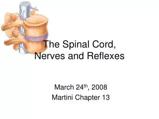
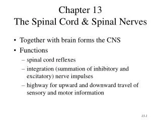
Chapter 13Spinal Cord, Spinal Nerves and Somatic Reflexes • Spinal cord • Spinal nerves • Somatic reflexes
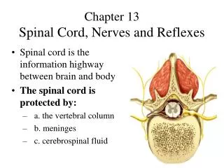
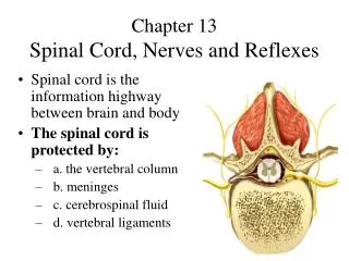
Overview of Spinal Cord • Information highway between brain and body • Extends through vertebral canal from foramen magnum to L1 • Each pair of spinal nerves receives sensory information and issues motor signals to muscles and glands • Spinal cord is a component of the Central Nervous System while the spinal nerves are part of the Peripheral Nervous System
Functions of the Spinal Cord • Conduction • bundles of fibers passing information up & down spinal cord • Locomotion • repetitive, coordinated actions of several muscle groups • central pattern generators are pools of neurons providing control of flexors and extensors (walking) • Reflexes • involuntary, stereotyped responses to stimuli (remove hand from hot stove) • involves brain, spinal cord and peripheral nerves
Anatomy of the Spinal Cord • Cylinder of nerve tissue within the vertebral canal (thick as a finger) • vertebral column grows faster so in an adult the spinal cord only extends to L1 • 31 pairs of spinal nerves arise from cervical, thoracic, lumbar and sacral regions of the cord • each cord segment gives rise to a pair of spinal nerves • Cervical & lumbar enlargements • Medullary cone is tapered tip of spinal cord • Cauda equinae is L2 to S5 nerve roots resemble horse’s tail
Meninges of the Spinal Cord • 3 Fibrous layers enclosing spinal cord • Dura mater • tough collagenous membrane surrounded by epidural space filled with fat and blood vessels • epidural anesthesia utilized during childbirth • Arachnoid mater • layer of simple squamous epithelium lining dura mater and loose mesh of fibers filled with CSF(creates subarachnoid space) • Pia mater • delicate membrane adherent to spinal cord • filium terminale and denticulate ligaments anchor the cord
Cross-Sectional Anatomy of the Spinal Cord • Central area of gray matter shaped like a butterfly and surrounded by white matter in 3 columns • Gray matter = neuron cell bodies with little myelin • White matter = myelinated axons
Gray Matter in the Spinal Cord • Pair of dorsal or posterior horns • dorsal root of spinal nerve is totally sensory fibers • Pair of ventral or anterior horns • ventral root of spinal nerve is totally motor fibers • Connected by gray commissure punctured by a central canal continuous above with 4th ventricle
White Matter in the Spinal Cord • White column = bundles of myelinated axons that carry signals up & down • Dorsal or posterior columns, lateral columns, and anterior columns • Each column is filled with named tracts (named fibers with a similar origin, destination & function)
Anatomy of a Nerve • A nerve is a bundle of nerve fibers (axons) • Epineurium covers nerves, perineurium surrounds a fascicle & endoneurium separates individual nerve fibers • Blood vessels penetrate only to the perineurium
Anatomy of Ganglia in the PNS • Cluster of neuron cell bodies in nerve in PNS • Dorsal root ganglion is sensory cell bodies • fibers pass through without synapsing
The Spinal Nerves • 31 pairs of spinal nerves (1st cervical above C1) • mixed nerves exiting at intervertebral foramen • Proximal branches • dorsal root is sensory input to spinal cord • ventral root is motor output of spinal cord • cauda equina is roots from L2 to C0 of the cord • Distal branches • dorsal ramus supplies dorsal body muscle & skin • ventral ramus to ventral skin & muscles & limbs • meningeal branch to meninges, vertebrae & ligaments
Branches of a Spinal Nerve Spinal nerves: 8 cervical, 12 thoracic, 5 lumbar, 5 sacral and 1 coccygeal. Each has dorsal and ventral ramus.
Nerve Plexuses • Ventral rami branch & anastomose repeatedly to form 5 nerve plexuses • cervical in the neck, C1 to C5 • supplies neck and phrenic nerve to the diaphragm • brachial in the armpit, C5 to T1 • supplies upper limb and some of shoulder & neck • lumbar in the low back, L1 to L4 • supplies abdominal wall, anterior thigh & genitalia • sacral in the pelvis, L4, L5 & S1 to S4 • supplies remainder of butt & lower limb • coccygeal, S4, S5 and C0
Structure of a Nerve Plexus • Notice the branching and merging of nerves in this example of a plexus
Nature of Somatic Reflexes • Quick, involuntary, stereotyped reactions of glands or muscle to sensory stimulation • automatic responses to sensory input that occur without our intent or often even our awareness • Functions by means of a somatic reflex arc • stimulation of somatic receptors • afferent fibers carry signal to dorsal horn of spinal cord • interneurons integrate the information • efferent fibers carry impulses to skeletal muscles • skeletal muscles respond
The Muscle Spindle • Sense organs that monitor the length of skeletal muscles (proprioceptors) = stretch receptors • respond to onset of stretch or prolonged stretch • 4 to 10 mm long modified skeletal muscle cells • intrafusal fibers that respond to gamma motor neurons & are wrapped with afferent fibers that respond to stretch
The Stretch (Myotatic) Reflex • When a muscle is stretched, it contracts & maintains increased tonus (stretch reflex) • helps maintain equilibrium & posture • head starts to tip forward as you fall asleep • muscles contract to raise the head • stabilize joints by balancing tension in extensors & flexors smoothing muscle actions • Very sudden muscle stretch causes tendon reflex • knee-jerk (patellar) reflex is monosynaptic reflex • testing somatic reflexes helps diagnose many diseases • Reciprocal inhibition prevents muscles from working against each other
Flexor Withdrawal Reflexes • Flexor(withdrawal) reflex occurs during withdrawal of foot from pain • polysynaptic reflex arc • neural circuitry in spinal cord controls sequence and duration of muscle contractions
Crossed Extensor Reflexes • Crossed extensor reflex maintains balance by extending other leg • intersegmental reflex extends up and down the spinal cord • contralateral reflex arcs explained by pain at one foot causes muscle contraction in other leg
Golgi Tendon Reflex • Proprioceptors in a tendon near its junction with a muscle -- 1mm long, encapsulated nerve bundle • Excessive tension on tendon inhibits motor neuron • muscle contraction decreased • Also functions when muscle contracts unevenly
Spinal Cord Trauma • 10-12,000 people/ year are paralyzed • 55% occur in traffic accidents • This damage poses risk of respiratory failure • Early symptoms are called spinal shock • Tissue damage at time of injury is followed by post-traumatic infarction