Navigating Lumbar Back Pain in Elite Athletes: Insights from Dr. Mark Gillett
Dr. Mark Gillett, Lead Physician at EIS West Midlands, explores the complexities of diagnosing and managing lumbar back pain in elite multi-sport athletes. Through case studies, he highlights the diagnostic dilemmas, particularly with imaging techniques like MRI and CT, and elaborates on extension-related pain mechanisms including discogenic issues, spondylolysis, and facet joint dysfunction. Emphasizing practical advice, he discusses the importance of history-taking, symptom tracking, and judicious use of imaging to enhance athletes' recovery while minimizing unnecessary radiation exposure.

Navigating Lumbar Back Pain in Elite Athletes: Insights from Dr. Mark Gillett
E N D
Presentation Transcript
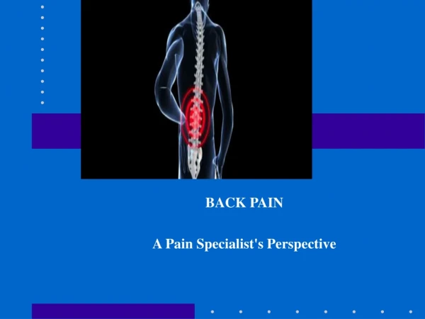
Lumbar Back Pain Dr Mark Gillett Lead Physician EIS West Midlands
Outline • Diagnostic categories • Imaging dilemmas in extension related back pain
Aims • Illustrate some of the problems regularly encountered by a sports physician working with an elite multi-sport population. • Put the literature to one side- what do I tell the athletes and coaches?
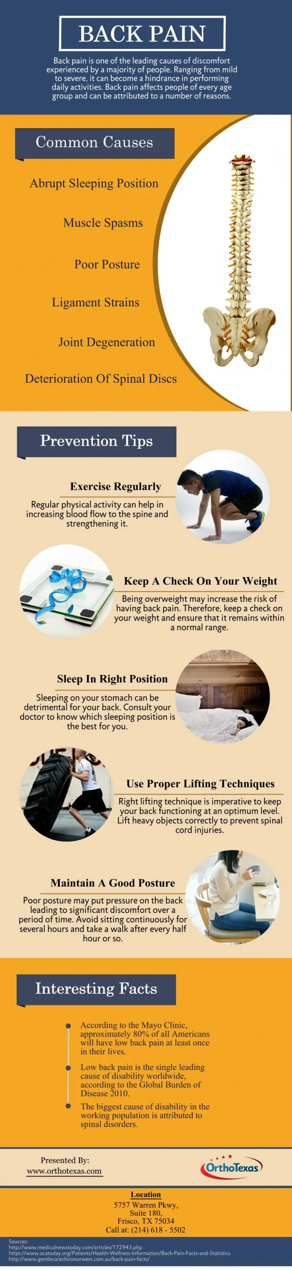
Types • Flexion related: discogenic pain • Extension related: pars interarticularis and facet joints
Recent Case Study • 22 year old heptathlete • 3 week history extension related back pain • Developed mumps • 10 day convalescence • Competed world indoor trials few days later • Saw track physician with back pain at meet
Recent Case Study • MRI scan- NAD (report only ) • Left lower facets and right mid-thoracic costo-transverse joint pain • 10 days manual therapy- no consistent improvement • Caudal epidural-no consistent improvement
Recent Case Study • Lumbar CT- avulsion fracture IAP of L1 at right L1-2 facet joint • Injection into fracture site- now pain free • Time from 1st consultation with me to pain free was 3 weeks
Problems Created • MRI entirely normal • Expectation of sport that all extension related pain with normal MRI will be followed by a CT
Extension Related Pain • Spondylolysis • Spondylolisthesis • Facet joint dysfunction
Diagnostic Strategy • History of extension related pain > few days • Pain and/or reduced range of motion in hyperextension • One legged hyperextension test (Stork test) • Facet joint loading
Investigation • MRI- lumbar spine to look for pars oedema or fluid in facet joints • Judicious use of CT • SPECT- not found it useful
Management Issues • Pars oedema useful sign on MRI but reliability of fracture identification is questionable • In a patient with a normal MRI- when is the correct time to progress to CT?
Juvenile Spondylolysis: a comparative analysis of CT, SPECT and MRICampbell et al Skeletal Radiol 2005 34:63-73 • General agreement good CT/SPECT vs MRI kappa:0.7866 • Discordance between groups when looking at stress reaction (1 in 11 case concordance) • Discordance between groups when looking at incomplete fractures ( 3/10 cases downgraded on MRI to bone stress, 3/10 upgraded on MRI to complete defects)
Take Home • MRI merely a screening tool to identify pars oedema • Athletes with pars oedema on MRI require a CT to detect a pars fracture
Facet Joints • Literature search for work examining sens/spec/PPV/NPV and inter-observer variation for MRI facet joint fluid • Nothing found • Most of literature devoted to similar analyses for disc degeneration
The prevalence of lumbar facet joint edema in patients with low back pain. Friedrich KM Skeletal Radiol. 2007Aug;36(8):755-60 • 145 consecutive MRI scans retrospectively reviewed by 2 musculoskeletal radiologists • Scan indication low back pain • Mean age 52.8 years • 14% showed bone marrow and soft tissue oedema at lumbar facets. All cases showed signs of OA. • L4-5 most common
Magnetic resonance imaging showed no signs of overuse or permanent injury to the lumbar sacral spine during a Special Forces training courseShachar Aharony MDaSpine J. 2007 Mar 2 • 10 soldiers underwent MRI of their lumbar sacral spines and right knees before and after the completion of a Navy Seals preparatory training lasting 14 weeks • Before the training, 7/10 spine MRIs were normal. 2 showed small L5–S1 disc bulges, 1 of these also had Scheuermann's disease. The 3rd soldier's MRI showed L1–L4 mild Scheuermann's disease. • Follow-up MRI showed no spinal changes
Demands • “Soldiers train wearing ceramic vests weighing 7 kg, carrying rifles weighing between 4 and 5.5 kg, and a pack up to 40% of their body weight while running and marching for distances up to 90 km. At the end of marches, teams of four soldiers walk and run over a hilly terrain, with a stretcher carrying a soldier with his full gear for up to 6 km. These soldiers are very motivated, have high pain tolerance, and are unlikely to seek medical care”
Conclusions • “These findings, along with the present study, suggest that young healthy trainees can do demanding activity in the short run without any evidence of damage to their lumbar sacral spines.”
Disagree • Doesn’t this suggest that MRI is not accurate enough to effectively screen out potentially significant spinal pathology in an athletic population?
E=Extension related back pain MRI Oedema No oedema CT Biomechanical analysis Time limited rehab Failed recovery Recovery CT
Advantages • Clear and transparent system for coaches and athletes • Utilises radiological investigations rationally and limits radiation exposure