Download
1 / 44
710 likes | 1.94k Vues
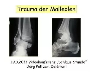
T r auma der Malleolen. 19.3.2013 Videokonferenz „Schlaue Stunde “ Jörg Peltzer, Delémont. Sprunggelenksfrakturen des Erwachsenen. Einführung. RX-Diagnostik. Klassifikationen/ Biomechanik. Operationstechniken. What`s New. Fazit. Einführung. Epidemiologie.

E N D
Trauma der Malleolen 19.3.2013 Videokonferenz „Schlaue Stunde“ Jörg Peltzer, Delémont
Sprunggelenksfrakturen des Erwachsenen Einführung RX-Diagnostik Klassifikationen/ Biomechanik Operationstechniken What`s New Fazit
Epidemiologie Zunahme der Inzidenz bei älteren Patienten um das 3-fache Häufigste Fraktur der unteren Extremität Risikofaktoren: erhöhter BMI, Raucheranamnese, Alkohol Isolierte Malleolarfraktur68 % Bimalleolarfraktur25 % Trimalleolarfraktur 7 % Offene Frakturen 2 %
OSG-Stabilitätskriterien Instabilität
Rx - Untersuchung ap, IR 30 ° lateral
pathologische Rx - Befunde Talussubluxation Talus Tilt Fibulaverkürzung
Klassifikation AO 44 A 44 B 44 C Infrasyndesmale Fraktur Transsyndesmale Fraktur Suprasyndesmale Fraktur
Klassifikation Lauge-Hansen Typ A Typ B Typ C Supination,Adduktion Supination, AR Pronation, Abduktion Pronation, AR
Klassifikationen medial Lateral Pronation Supination
Typ A Frakturen Supination, Adduktion
Push off Pull off
Typ A Frakturen Supination ,Adduktion
Typ B Frakturen Supination, Aussenrotation
Volkmann Dreieck Wann fixieren? 1/3 oder 1/4 Regel
isolierte Fraktur Malleolus int. Robert Adams
Typ C Frakturen pre-op post-op
Sichere Zone ½ ½ posterior anterior Schrauben posteriorColliculusanterior Risiko Verletzung Tib.post. Sehne Femino JE, JBJS 89A (1) 133, 2007
Frakturtypen 1 2 3 1 Vertikal 2 Transversal 3 Avulsion
Frakturtypen/Versorgung 1 Vertikal 2 Transversal 3 Avulsion
bikortikale Schraubenlage ist unikortikaler klar überlegen Zuggurtung Stahl Fibre wire unikortikal bikortikal Orthopedics, Aug 2011, 34 (8); 349-355
bei vertikaler Fraktur divergente Schraubenlage am stabilsten parallel konvergent divergent J Trauma 72 (3): 751-54; 2012
What`s New ? MIPO Implantate Nachbehandlung
Nachbehandlung 70% Vollbelastung 30 % Teilbelastung
Fazit Read theFracture, Pronation oder Supination Verstehen der Biomechanik, Rotation / Translation Erkennung der Verletzungen:Knochen und Bänder Analyse der Instabilität Entscheid ihrer Implantate Volkmann: Fixation bei ¼ oder 1/3 Med.Malleolus:vertical,transversal Optimale Wahl des Operationszeitpunktes Gewebeschonende OP-Technik mit der FX und dem Patienten angepassten Implantaten wenn immer möglich funktionelle Nachbehandlung