Download
1 / 1
10 likes | 217 Vues
Stability Enhancement of DNA Nanogrids via Streptavidin-Biotin Complexation Dawn A. Bonnell, University of Pennsylvania, DMR 0425780.

E N D
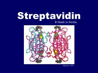
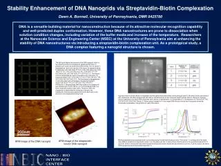
Stability Enhancement of DNA Nanogrids via Streptavidin-Biotin Complexation Dawn A. Bonnell, University of Pennsylvania, DMR 0425780 DNA is a versatile building material for nanoconstruction because of its attractive molecular recognition capability and well-predicted duplex conformation. However, these DNA nanostructures are prone to dissociation when solution condition changes, including variation of the buffer media and increase of the temperature. Researchers at the Nanoscale Science and Engineering Center (NSEC) at the University of Pennsylvania aim at enhancing the stability of DNA nanostructures via introducing a streptavidin-biotin complexation unit. As a prototypical study, a DNA complex featuring a nanogrid structure is chosen. The left figure depicts the structure of the DNA nanogrid, which is two-dimensional and is composed of repeating DNA tiles. To prepare such a grid, we need three DNA strands: the first strand is designated as i and is the red one in the figure, its sequence being 5’-AGG CAC CAT CGT AGG TTT TCT TGC CAG GCA CCA TCG TAG GTT TTC TTG CCA GGC ACC ATC GTA GGT TTT CTT GCC AGG CAC CAT CGT AGG TTT TCT TGC C-3’. The second strand is designated as ii and is the yellow one in the figure, its sequence being 5’-ACT ATG CAA CCT GCC TGG CAA GCC TAC GAT GGA CAC GGT AAC G-3’. The third strand is designated as iii and is the green one in this figure, its sequence being 5’-/biotin/CGC GCG TTA CCG TGT GGT TGC ATA GTC ATG/biotin/-3’. Bridging these DNA tiles relies on the designed 5’-end (CGCG) and 3’-end (CATG) in iii. In this bridging mode, four biotin molecules reside at each splint. Therefore, when the streptavidin is added after the formation of the grid, two streptavidin molecules are expected to bind to each splint via a two-biotin-one-streptavidin binding mode. A gel test result is shown above. It is apparent that the splints dissociated when running the gel (lane 3). But when biotins were bound with streptavidin, the DNA nanogrid was still held up and behaved as a high molecular weight species (lane 4). Being an important control experiment, one more DNA strand was also used as a replacement of iii. Designated as iv, its sequence is 5’-/biotin/CGT TAC CGT GTG GTT GCA TAG T/biotin/-3’. When using iv instead of iii, only single DNA tiles are formed, and its property should be intrinsically comparable to the grid after its splint dissociation. Melting temperature measurement result, and corresponding first derivative analysis result. The curves 1- 4 correspond to the same oligo addition conditions as specified in lanes 1-4 in the above gel test. It clearly shows that the splints in the grid dissociated at 52 oC (curve 3), while no dissociation was observed at this temperature when streptavidin bound biotins (curve 4). In all four cases, the whole DNA nanogrid structure dissociated beyond 60 oC. AFM image of the streptavidin-bound DNA nanogrid AFM image of the DNA nanogrid