Download
1 / 37
370 likes | 506 Vues
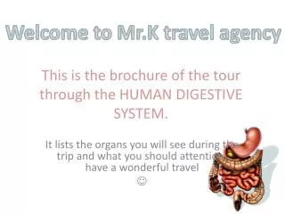
Welcome to the luxury tour Through the digestive system. By Silvia Chen. I’m Qoo . The tourist of the trip Let’s begin !! . Map of our trip. Alimentary canal. First scenic spot: Oral Cavity.

E N D
Welcome to the luxury tour Through the digestive system By Silvia Chen
I’m Qoo. The tourist of the trip Let’s begin !!
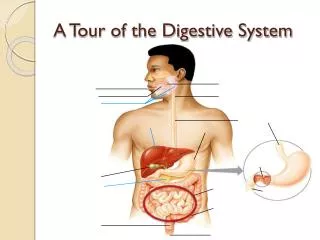
Map of our trip Alimentary canal
First scenic spot: Oral Cavity Here we are in the mouth. The fancy term that leaps to the eyes names oral cavity. Let enter it! • Tongue • Teeth • Salivary glands 4.Pharynx 5.Epiglottis 6.Esophagus
Tongue The place we stand on is called tongue. It’s soft and pink-colored. It is the primary organ of taste, as much of the upper surface of the tongue is covered n papillae and taste buds. It’s It is sensitive and kept moist by saliva, and is richly supplied with nerves and blood vessels.
Teeth The white, little stuffs are named teeth. It starts the process of physical digestion by breaking down food into smaller pieces.(biting) After that, the chewed food is pushed to the pharynx. Teeth decay
Teeth decay Tooth decay disease is caused by specific types of bacteria that produce acid in the presence of fermentable carbohydrates such as sucrose, fructose, and glucose. The mineral content of teeth is sensitive to increases in acidity from the production of lactic acid. In order to protect teeth, Brush our teeth more regularly, Eat less sweet, And go to see the dentist in a regular time
Salivary glands The fluid spilled over our head is called saliva, which is produced by salivary glands. Saliva is used to moistens food, and contains enzymes that begin the chemical digestion of carbohydrates. 3 types of salivary glands: Sublingual gland(beneath the tongue) Sub maxillary gland(under the mouth) Parotid gland(in front of the ears)
Enzyme in saliva Salivary Amylase: It is used in the digestion of carbohydrate and acts on starch t break it into many molecules of maltose
Pharynx Continue walking, we can see little stuff which is named pharynx. It’ sin the back of the throat where oral and nasal cavities join. Tongue pushes chewed food to pharynx that swallowing occurs.
Epiglottis Look down to the deep passageway, the flap of tissue like a little door names epiglottis. It closes off the opening of the trachea when swallowing food. It also keeps food from entering the air passage.
Esophagus Here the deep and dark passageway under us is called esophagus. It is the tube leading from the pharynx to the stomach. Food moves down the it due to the process of peristalsis. Next, we are going to experiencing the interesting “peristalsis”! Let’s jump down!
Peristalsis, peristalsis> < Let’s go down with a rhythmical contraction of the muscles that lines the esophageal wall pushes food down. > < DOWN DOWN DOWN
Second scenic spot: The abdominal cavity 1.Stomach 2. Duodenum 3. Gall bladder 4.Liver 5.Small intestine 6.Large intestine 7.Appendix 8.Rectum 9.anus
Here we are in a J-shaped organ, you see, named stomach. We are standing in the cardiac sphincter, the top of the stomach. The band of muscle behind us, like a door closes off the top of the stomach in order to keep stomach acid from entering the esophagus. Food is stored and churned in it! Churning helps physical digestion of food and results in a product called acid chyme. Stomach
Enzymes in the stomach Continue walking,it’s clear to see the chemical digestion of protein begins. But what helps it? The answer is pepsinwhich is the product that pepsinogen and HCI. These two things are produced by the gastric glands, which is stimulated by the gastrin, entering the bloodstream, in the upper part of stomach. HCI(Acidic pH), in front of us, can be seen clearly in a big “pool”. However…
Something bad happens The wall of the stomach is covered with mucous that is produced to protect the stomach lining(HCI can burn the lining of the stomach). However, if a portion of the stomach does get burned, ulcer occurs! Peptic ulcers may even lead to bleeding or perforation, emergency situation!
Pyloric Sphincter Pass through the HCI pool, we can see another door that seems the same as cardiac sphincter names pyloric sphincter. It’s a band of muscle which closes off the bottom of the stomach in order to prevent HCI from entering the duodenum and allows small amounts of chyme to enter the intestine.
Duodenum After we exits the stomach, we walk into a 10-inche long gallery called duodenum. We can see various fluids spilled over our head! What are they? The answer will be revealed soon. First let me introduce the duodenum. It’s the place that chemical digestion of all 3 food groups(proteins, fats, carbohydrates) with the help of the enzymes form the pancreas and bile from the gall bladder.
Hormones produce by duodenal wall -Secretin: stimulate the release of pancreatic juice from the pancreas and is stimulated by HCI. (in response to the release of pancreatic juice) -cholecystokinin (cck): stimulates the release of pancreatic juice from pancreas and is stimulated by partially digested protein and fat. . (in response to the release of pancreatic juice) Both of them then enter the blood stream.
Pancreas Open our umbrella and go to find the resource of the fluids! Here they are! They come from a place named pancreas that produce digestive enzymes and sodium bicarbonate, which is used to neutralize the acid chyme and pH comes to neutral pH. It is called both an exocrine organ that produces enzymatic substances and an endocrine organ that produces hormones. It provides huge contribution, right!
Enzyme produces by pancreas -Lipase: breaks down fat droplets into glycerol and 3 fatty acids Lipase Fat droplets + Water Glycerol+3 fatty acids -Pancreas Amylase: acts on starch to convert it to maltose. - Trypsin: breakdown proteins to peptides trypsin peptides Protein + water
Hormones in pancreas -Insulin: secreted when blood sugar concentration is high and causes liver and muscles to take up and store excess glucose as glycogen. It also promotes synthesis of protein and fats. Insulin Low blood sugar High blood sugar Glucagon -Glucagon: secreted when blood sugar concentration is low and causes liver and muscles to break down glycogen into glucose. It also stops protein and fat synthesis. (raise blood sugar level)
Ball gladder Let’s return to duodenum. The little ball-shaped stuff above us is called ball gladder. It stores bile. Bile is used to emulsify fats, which means to break fat down into small fat droplets in the duodenum. BILE IS NOT AN ENZYME!
Liver The red area above the gall bladder is called liver, which is an essential organ. I’m going to introduce some of its functions: -It produces bile. -It destroys old red blood cells and converts hemoglobin to a product in bile. -Store glucose as glycogen after eating, and break down glycogen to glucose between eating to help maintain glucose level of blood. -It produces urea from the breakdown of amino acids(deamination) -make blood protein from amino acids. -Helps to detoxifies the blood by removing poisonous substances and metabolizing them.
Small intestine Pass through the gallery, we enter the mot amazing place,I think, called small intestine. Why it is so amazing? Because it’s 7 meters long!!! It folds and convolutes to create more surface area. Final food digestion occurs in it. Look at the wall of the small intestine, there are countless hair-like things called villus, which are sites of absorption of nutrients. Each villi has thinner microvilli, small blood vessels and a small lymph called lacteal, which absorbs fluids and returns it to the veins. More about villus
Villus Villus capillaries also collect amino acids and simple sugars taken up by the villi into the blood stream. Villi are specialized for absorption in the small intestine as they have a thin wall, about one cell thick, which enables a shorter diffusion path. They have a large surface area so there will be more efficient absorption of fatty acids and glycerol into the blood stream. They have a rich blood supply to keep a concentration gradient. -small, finger-like projections that protrude from the epithelial lining of the intestinal wall. -approximately 0.5-1.6 (mm) in length and has many microvilli (singular: microvillus), each of which are much smaller than a single villus. The intestines villi is approximately around 200m2
Enzymes in small intestine -Maltase: converts maltose to glucose maltase Maltose + water glucose -peptidase: break down peptides into amino acids peptidase Peptides + Water Amino acids -nuclease: (also in pancreas) break down RNA and DNA into nucleotides
Walk.. Walk.. Walk… Oh, it’s too long!!!
Large intestine(colon) After a long time… Now we are standing at the end of small intestine and the beginning of the colon called caecum(belong to large intestine). The colon is responsible for absorption of water from undigested food and some vitamin. It can be divided into 4 parts: ascending colon, transverse colon, descending colon and sigmoid colon.
E. Coli bacteria The little fish-like stuffs called e.coli bacteria exist in colon to consume any substances that were not digested earlier It’s amusing that when the bacteria breaks down these substances, they give off odorous molecules that cause the characteristic odor of feces or “passing gas”. E. coli helps to metabolize what the bodies are unable to metabolize, thereby providing us with a vital service.
Appendix Look at the little tail over there at the junction of the small and large intestine names appendix. Now, the function of it is still a mystery.
Rectum Here is the end of the large intestine and it’s a enlarged portion named rectum. It stores undigested food temporarily. Oh the smell is awful here! Let’s get out of here! Jump though the anus, which is bands of muscle that allow undigested wastes(poop) to exit the body! I can even smell the fresh air!
Pooping Oh my gosh! I go with feces which are the undigested food and waste and smelly out of the body!! All contents of food that cannot be digested exit the body by the process named elimination, defecation or pooping. With the process, our trip ends as well! I had to take a shower first!!!
Qoo knowledge window: -Digestion: The organic process by which food is converted into substances that can be absorbed into the body. -The term called Physical digestion is physical breakdown of food into smaller pieces and increasing the surface area so enzyme can work on them. -The term called chemical digestion is chemical breakdown of food using enzyme.
Reference Chemical Digestion, Absorption PPT, TanghuasWikispace Tooth decay: http://en.wikipedia.org/wiki/Dental_caries Tongue: http://en.wikipedia.org/wiki/Tongue Villus: http://en.wikipedia.org/wiki/Intestinal_villus
Our trip ends in a satisfactory way! I have an enjoyable time with you! I hope to see you again, my friends! Bye~