Ch 16: Blood
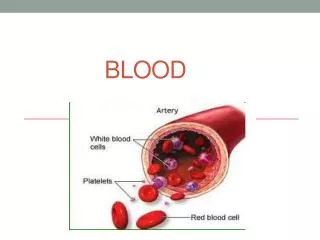
Ch 16: Blood. Plasma and Cellular Elements of Blood Hematopoiesis RBC Physiology Coagulation. Blood = connective tissue. Extracellular matrix:. Specialized cells:. Fig 16-1. Plasma. Blood Components Overview. Blood. 20-40%. Total WBC: 4,000 - 11,000. 2-8%. Cellular Elements.

Ch 16: Blood
E N D
Presentation Transcript
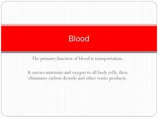
Ch 16: Blood Plasma and Cellular Elements of Blood Hematopoiesis RBC Physiology Coagulation
Blood = connective tissue Extracellular matrix: Specialized cells: Fig 16-1
Plasma Blood Components Overview Blood 20-40% Total WBC: 4,000 - 11,000 2-8% Cellular Elements 50-70% 1- 4% Fig 16-1/3
Red Blood Cells O2 Fig 16-5
Hem(at)opoiesis = Blood Cell Formation • Few uncommitted stem cells in red bone marrow throughout life time (Fig 16-2) • Controlled by cytokines. Examples: • Erythropoietin • CSFs and ILs: e.g. M-CSF, IL-3 (= multi CSF) • Thrombopoietin • Leukemia vs. leukocytosis vs. leukopenia
Controlled by ____________,specifically CSFs and ILs Compare to Fig 16-2
EPO Regulates RBC Production • “Hormone” synthesized by kidneys in response to hypoxemia • EPO gene cloned in 1985 Recombinant EPO now available (Epogen, Procrit) • Use in therapy, abuse in sport
Erythropoiesis EPO release Mitotic rate Tissue O2 RBC bag of Hbfor carrying O2 lifespan ~ 120 days source of ATP for RBC? Maturation speed Tissue O2 Reticulocytes enter circulation
Hemoglobin (Hb) • Requires iron (Fe) + Vit. B12 (cobalamin) p.698/Ch21 • Quaternary protein structure ? • Reversible binding between Fe & O2 • CO: a toxic gas (not in book) • Bilirubin to bile. Hyperbilirubinemia • HbA vs. HbF
Hb Structure Porphyrin ring with Fe in center How many O2 can 1Hb carry?
RBC Disorders • Polycythemia vera (PCV ~ 60-70%) • Anemias(O2 carrying capacity too low) • Hemorrhagic anemia Fe deficiency anemia • Hemolytic anemia, due to genetic diseases (e.g. Hereditary spherocytosis) or infections • Pernicious anemia • Renal anemia
Sickle Cell Anemia 1st genetic illness traced to a specific mutation: DNA:CAC CTC aa: glutamic acid valine (aa #6 of 146) HbA HbS crystallizes under low oxygen conditions
Platelets = Thrombocytes • Megakaryocytes (MKs) are polyploid. Mechanism? • MK produces ~ 4,000 platelets which live an average of 10 days. • Platelets contain gra-nules filled with clotting proteins & cytokines • Activated when blood vessel wall damaged
Hemostasis = Opposite of hemorrhage stops bleeding Too little hemostasis too much bleeding Too much hemostasis thrombi / emboli Three major steps: • Vasoconstriction • Platelet plug(temporary blockage of hole) • Coagulation (clot formation seals hole until tissues repaired)
Steps of Hemostasis Vessel damage exposes collagen fibers Platelets adhere to collagen & release factors local vasoconstriction & platelet aggregation decreased blood flow platelet plug formation + feedback loop Fig 16-11
Platelet Plug Formation Platelet activating factor (PAF)
Steps of Hemostasis cont. Two coagulation pathways converge onto common pathway • Intrinsic Pathway. Collagen exposure. All necessary factors present in blood. Slower. • Extrinsic Pathway. Uses TF released by injured cells and a shortcut. • Usually both pathways are triggered by same tissue damaging events. Fig 16-12
The Coagulation Cascade “Cascade” is complicated network! Numbering of coagulation factors according to time of discovery Fig 16-12
Common Coagulation Pathway Intrinsic pathway Extrinsic pathway Active factor X Prothrombin thrombin fibrinogen fibrin reinforces platelet plug clot
Structure of Blood Clot Plasmin, trapped in clot, will dissolve clot by fibrinolysis Clot formation limited to area of injury: Intact endothelial cells release anticoagulants (heparin, antithrombin III, protein C). SEM x 4625
Clot Busters & Anticoagulants Dissolve inappropriate clots Enhance fibrinolysis Examples:Urokinase, Streptokinase & t-PA Prevent coagulation by blocking one or more steps in fibrin forming cascade Inhibit platelet adhesion plug prevention Examples:
Blood Doping the end