Blood Vessels: The Vascular System
Blood Vessels: The Vascular System. Transport blood to the tissues and back Carry blood away from the heart Arteries Arterioles Exchanges between tissues and blood Capillary beds Return blood toward the heart Venules Veins. Blood Vessels: The Vascular System. Figure 11.9a.


Blood Vessels: The Vascular System
E N D
Presentation Transcript
Blood Vessels: The Vascular System • Transport blood to the tissues and back • Carry blood away from the heart • Arteries • Arterioles • Exchanges between tissues and blood • Capillary beds • Return blood toward the heart • Venules • Veins
Blood Vessels: The Vascular System Figure 11.9a
Blood Vessels: Microscopic Anatomy • :; • : • .
Blood Vessels: The Vascular System Figure 11.9b
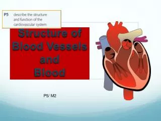
Structure of Capillary Walls Capillary walls are essentially just the tunica intima (endothelium plus the basement membrane); thus, they are exceedingly thin.
Differences Between Blood Vessels • Walls of arteries are the thickest • Lumens of veins are larger • Larger veins have valves to prevent backflow • Skeletal muscle “milks” blood in veins toward the heart • Walls of capillaries are only one cell layer thick to allow for exchanges between blood and tissue
Blood Vessels: The Vascular System Arteries are much closer to the pumping action of the heart and must be able to withstand the pressure fluctuations at such locations. Veins, on the distal side of the capillary beds of the tissues, are essentially low-pressure vessels that need less strength/support/ elasticity than do arteries. Figure 11.9a
Skeletal Muscles and Valves The presence of valves, the milking action of skeletal muscles against the veins as the muscles contract, the respiratory pump (pressure changes in the thorax during breathing) Figure 11.10
Breathing in pulls blood into right atrium from vena cava • the “thoracic pump” which generates negative pressure in the lungs during inhalation causing blood to rush through the vena cava and right heart filling the dense The thoracic pump draws blood from the extremities on inhalation and sends blood throughout the body on exhalationpulmonary capillary bed
Pulmonary arteries carry oxygen-poor blood and pulmonary veins carry oxygen-rich blood. • Umbilical arteries carry oxygen-poor blood from the fetus and the umbilical vein carries the most oxygen-rich blood to the fetus.
Movement of Blood Through Vessels • Most arterial blood is pumped by the heart • Veins use the milking action of muscles to help move blood
Capillary Beds • Capillary beds consist of two types of vessels • Vascular shunt—vessel directly connecting an arteriole to a venule • True capillaries—exchange vessels • Oxygen and nutrients cross to cells • Carbon dioxide and metabolic waste products cross into blood
Capillary Beds Figure 11.11a
Capillary Beds Figure 11.11b
Major Arteries of System Circulation • Aorta • Largest artery in the body • Leaves from the left ventricle of the heart • Regions • Ascending aorta—leaves the left ventricle • Aortic arch—arches to the left • Thoracic aorta—travels downward through the thorax • Abdominal aorta—passes through the diaphragm into the abdominopelvic cavity
Major Arteries of System Circulation • Arterial branches of the ascending aorta • Right and left coronary arteries serve the heart
The Heart Figure 11.2a
Major Arteries of Systemic Circulation • Arterial branches of the aortia arch (BCS) • Brachiocephalic trunk splits into the • Right common carotid artery • Right subclavian artery • Left common carotid artery splits into the • Left internal and external carotid arteries • Left subclavian artery branches into the • Vertebral artery • In the axilla, the subclavian artery becomes the axillary artery brachial artery radial and ulnar arteries
Major Arteries of Systemic Circulation • Arterial branches of the thoracic aorta • Intercostal arteries supply the muscles of the thorax wall • Other branches of the thoracic aorta supply the • Lungs (bronchial arteries) • Esophagus (esophageal arteries) • Diaphragm (phrenic arteries)
Major Arteries of Systemic Circulation • Arterial branches of the abdominal aorta • Celiac trunk is the first branch of the abdominal aorta. Three branches are • Left gastric artery (stomach) • Splenic artery (spleen) • Common hepatic artery (liver) • Superior mesenteric artery supplies most of the small intestine and first half of the large intestine
Major Arteries of Systemic Circulation • Arterial branches of the abdominal aorta • Left and right renal arteries (kidney) • Left and right gonadal arteries • Ovarian arteries in females serve the ovaries • Testicular arteries in males serve the testes • Lumbar arteries serve muscles of the abdomen and trunk
Major Arteries of Systemic Circulation • Arterial branches of the abdominal aorta • Inferior mesenteric artery serves the second half of the large intestine • Left and right common iliac arteries are the final branches of the aorta • Internal iliac arteries serve the pelvic organs • External iliac arteries enter the thigh femoral artery popliteal artery anterior and posterior tibial arteries
Left ventricle to ascending aorta aortic arch brachiocephalic artery subclavian artery axillary artery brachial artery radial (or ulnar) artery • capillary network of wrist • radial (or ulnar) vein brachial vein axillary vein subclavian vein right brachiocephalic vein superior vena cava right atrium of the heart. Figure 11.12
Left ventricle ascending aorta aortic arch descending aorta right common iliac artery external iliac artery femoral artery poplitealartery anterior tibial artery dorsalispedis artery • capillary network • anterior tibial vein popliteal vein femoral vein external iliac vein common iliac vein inferior vena cava right atrium of the heart. Figure 11.12
Major Veins of Systemic Circulation • Superior and inferior vena cava enter the right atrium of the heart • Superior vena cava drains the head and arms • Inferior vena cava drains the lower body
The Heart Figure 11.2b
Major Veins of Systemic Circulation • Veins draining into the superior vena cava • Radial and ulnar veins brachial vein axillary vein • These veins drain the arms • Cephalic vein drains the lateral aspect of the arm and empties into the axillary vein • Basilic vein drains the medial aspect of the arm and empties into the brachial vein • Basilic and cephalic veins are jointed at the median cubital vein (elbow area)
Major Veins of Systemic Circulation • Veins draining into the superior vena cava • Subclavian vein receives • Venous blood from the arm via the axillary vein • Venous blood from skin and muscles via external jugular vein • Vertebral vein drains the posterior part of the head • Internal jugular vein drains the dural sinuses of the brain
Major Veins of Systemic Circulation • Veins draining into the superior vena cava • Left and right brachiocephalic veins receive venous blood from the • Subclavian veins • Vertebral veins • Internal jugular veins • Brachiocephalic veins join to form the superior vena cava right atrium of heart • Azygous vein drains the thorax
Major Veins of Systemic Circulation • Veins draining into the inferior vena cava • Anterior and posterior tibial veins and fibial veins drain the legs • Posterior tibial vein popliteal vein femoral vein external iliac vein • Great saphenous veins (longest veins of the body) receive superficial drainage of the legs • Each common iliac vein (left and right) is formed by the union of the internal and external iliac vein on its own side
Major Veins of Systemic Circulation • Veins draining into the inferior vena cava • Right gonadal vein drains the right ovary in females and right testicle in males • Left gonadal vein empties into the left renal vein • Left and right renal veins drain the kidneys • Hepatic portal vein drains the digestive organs and travels through the liver before it enters systemic circulation
Major Veins of Systemic Circulation • Veins draining into the inferior vena cava • Left and right hepatic veins drain the liver
Major Veins of Systemic Circulation Figure 11.13
Arterial Supply of the Brain • Internal carotid arteries divide into • Anterior and middle cerebral arteries • These arteries supply most of the cerebrum • Vertebral arteries join once within the skull to form the basilar artery • Basilar artery serves the brain stem and cerebellum
Arterial Supply of the Brain • Posterior cerebral arteries form from the division of the basilar artery • These arteries supply the posterior cerebrum
Circle of Willis • Anterior and posterior blood supplies are united by small communicating arterial branches • Result—complete circle of connecting blood vessels called cerebral arterial circle or circle of Willis
Arterial Supply of the Brain Figure 11.14
Fetal Circulation • Fetus receives exchanges of gases, nutrients, and wastes through the placenta • Umbilical cord contains three vessels • Umbilical vein—carries blood rich in nutrients and oxygen to the fetus • Umbilical arteries (2)—carry carbon dioxide and debris-laden blood from fetus to placenta
Fetal Circulation • Blood flow bypasses the liver through the ductus venosus and enters the inferior vena cava right atrium of heart • Blood flow bypasses the lungs • Blood entering right atrium is shunted directly into the left atrium through the foramen ovale • Ductus arteriosus connects the aorta and pulmonary trunk (becomes ligamentum arteriosum at birth)
Fetal Circulation Figure 11.15
Hepatic Portal Circulation • Veins of hepatic portal circulation drain • Digestive organs • Spleen • Pancreas • Hepatic portal vein carries this blood to the liver • Liver helps maintain proper glucose, fat, and protein concentrations in blood
Hepatic Portal Circulation • Major vessels of hepatic portal circulation • Inferior and superior mesenteric veins • Splenic vein • Left gastric vein
Hepatic Portal Circulation Figure 11.16
Hepatic Portal Circulation Figure 11.17
Pulse • Pulse • Pressure wave of blood • Monitored at “pressure points” in arteries where pulse is easily palpated • Pulse averages 70–76 beats per minute at rest
Pulse Figure 11.18
Blood Pressure • Measurements by health professionals are made on the pressure in large arteries • Systolic—pressure at the peak of ventricular contraction • Diastolic—pressure when ventricles relax • Write systolic pressure first and diastolic last (120/80 mm Hg) • Pressure in blood vessels decreases as distance from the heart increases