Download

1 / 39
400 likes | 490 Vues
Learn technical considerations for mobile X-ray units used in large animal imaging, from safety to common developmental conditions like OCD and ALD. Discover radiographic findings, fractures, DJD, Navicular Disease, Physitis, Osteomyelitis, and Laminitis.

E N D
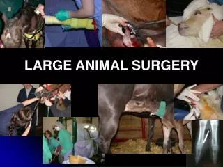
Introduction to Large AnimalMusculoskeletal Imaging Dr. LeeAnn Pack
Technical Considerations • Usually mobile units used • Min-X • Bowie • Thorax, shoulders etc – need bigger machine • FFD is shorter usually 30 inches • Should check with the measuring tape • Collimation
Safety • Barns • People need to be out of barn • Pregnant? • Radiation Protection • Lead aprons, gloves • Take care of them in the truck – no folding • Cassette holder, wooden blocks • Sedation
The Radiographs • Correct Naming of views • Correct Labeling of films • View (R, L, DMPLO etc) • Label correct (patient name etc) • Correct Viewing of films and naming of structures seen • Legality
Common Developmental Conditions • OCD • Cartilage fails to ossify • Free fragment or flap • Radiographic findings: • Flattening of the articular surface • Mineralized fragment or flap • Subchondral lucency/sclerosis • Secondary DJD • Often bilateral
Common Developmental Conditions • OCD – Subchondral Bone Cyst • Articular cartilage is stopped but then returns – area of cartilage gets trapped and will be seen as a lucency in the subchondral bone • Secondary DJD can develop • Often bilateral
Angular Limb Deformity • Medial or lateral deviation of the limb • Valgus = lateral • Varus = medial • May be d/t • abnormal cuboidal bones • Uneven physeal growth • Ligamentous laxity
ALD • Usually can tell just looking at the foal • Shoot DP rads including joint above and joint below • Then can draw lines to tell exactly where the deviation originates • Windswept • Repair
Fractures • 3rd carpal bone • Radial carpal bone • Kissing lesions • P3 fractures • Apical sesamoid fractures • Splint bone fractures
Degenerative Joint Disease • Most common joint disease in horses • Primary - idiopathic • Secondary – most common • Repetitive motion/movements • Running to the left • Common in Tarsus, Carpus, “Fetlocks” and IP joints
DJD • Radiographic findings: • Thickening of joint capsule • Narrowed joint space • New bone production on articular margins (osteophytes) • Subchondral bone sclerosis • Ankylosis of joints
Degenerative Joint Disease • Tarsus - Spavin • Carpus • Stifle • “Fetlocks” • Distal – IP joints • Ring bone • High – DJD of proximal IPJ • Low – DJD of distal IPJ
Navicular Disease • Chronic progressive disease that affects the distal sesamoid bone • Usually bilateral • Fore limb more than hind limb • Quarter horses with some TB’s • Navicular disease should not be used as a radiographic diagnosis
Navicular Disease • Radiographic findings • Synovial invaginations enlarged and too numerous • Medullary sclerosis • Lollipops
Physitis • Related to nutrition? • Young rapidly growing foals • Distal radius, distal tibia, distal 3rd metacarpal and metatarsal bones • Multiple physes usually involved • Radiographic signs • Metaphyseal flaring with sclerosis next to physis • Cortical thickness asymmetry – widened irregular physis
Osteomyelitis • Foals • Hematogenous spread • Umbilicus, lungs, liver – bacterial showering • Adults • Sequestrum can occur • Usually due to trauma • Radiographic findings • Multiple bones? Single bone? Lysis near physis – septic joint
Laminitis • Inflammation of the laminae of the foot • Cause? • Front feet usually – often bilateral • Saw horse stance • Lack of parallelism between the dorsal aspect of the hoof wall and dorsal aspect of P3 – ST of P3 thick • 18mm • How to ID on rads – rotation, sinking