Download
1 / 69
690 likes | 877 Vues
The Nervous System. E. McIntyre Biology 12. Nervous System. Central Nervous System. Peripheral Nervous System. Somatic Nerves. Autonomic Nerves. Spinal Cord. Brain. Sensory. Motor. Sympathetic. Parasympathetic. Forebrain. Midbrain. Hindbrain. The Four Lobes…. Cerebrum.

E N D
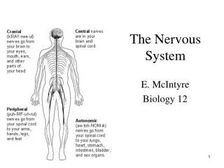
The Nervous System E. McIntyre Biology 12
Nervous System Central Nervous System Peripheral Nervous System Somatic Nerves Autonomic Nerves Spinal Cord Brain Sensory Motor Sympathetic Parasympathetic Forebrain Midbrain Hindbrain The Four Lobes… Cerebrum Olfactory Lobes Thalamus Pons Medulla Oblongata Corpus Callosum Cerebellum Hypothalamus
CNS & PNS • CNS – coordinating centre for information coming in and going out (brain and spinal cord) • PNS – carries information between organs of the body and the CNS much more later… http://diabetes.niddk.nih.gov/dm/pubs/complications_nerves/index.htm
Central Nervous System (CNS) • comprised of brain and spinal cord • coordinating centre for information coming in and going out • surrounded by bone-skull and vertebrae. • fluid (cerebrospinal fluid) and fluid (meninges) insulate the brain and spinal cord. http://diabetes.niddk.nih.gov/dm/pubs/complications_nerves/index.htm
…Central Nervous System (CNS) • The brain is composed of three main parts: • Cerebrum (seat of consciousness) • Cerebellum: muscle coordination and maintains normal muscle tone and posture. The cerebellum coordinates balance • medulla oblongata regulation of heartbeat, breathing, vasoconstriction (blood pressure), and reflex centers for vomiting, coughing, sneezing, swallowing, and hiccupping
Peripheral Nervous System (PNS) • carries information between organs of the body and the CNS
Somatic Nerves • Somatic Nerves lie outside the CNS and therefore are part of the PNS.
Autonomic Nerves • Controls the internal organs • Sympathetic: turns ‘on’ systems • OPENS eyes to allow more light in • DIALATES bronchioles in lungs • INCREASES heart rate • Parasympathetic: turns ‘off’ systems • CLOSES eyes to reduce light coming in • CONSTRICTS bronchioles in lungs
Types of Nerve Cells There are two types of nerve cells • Glial cells • Non-conducting cells: structure/support and metabolism of nerve cells • Neurons • Nerve cells that conduct electrical signalscalled nerve impulses. • carries impulses in only ONE direction • Basic functional unit of the Nervous System
Types of Neurons Somatic nerves control skeletal muscle, bones, skin • Sensory (AFFERENT) – relay information from sense organs (eyes, nose, mouth etc) to CNS. • Interneurons – Connect Sensory and Motor neurons and carry impulses between them. They are found entirely within the Central Nervous System. • Motor neurons (EFFERENT) – relay information from CNS to the effectors (muscles, organs, glands)
A neuron has three main parts Body: largest part, contains the nucleus and much of cytoplasm, most of the metabolic activity of the cell, including the generation of ATP and synthesis of protein. Dendrites: Short branch extensions spreading out from the cell body. Dendrites Receive STIMULUS (Action Potentials) and carry IMPULSES from the ENVIRONMENT or from other NEURONS AND carry them toward cell body. Axon: A Long Fiber that CARRIES IMPULSES AWAY FROM THE CELL BODY. Each neuron has only ONE AXON. The axon ends in a series of small swellings called AXON TERMINALS. Structure of the Neuron Structure of Neuron
Neurons may have dozens or even hundreds of dendrites but usually only one axon. Nodes of Ranvier: gaps between sections of myelin sheath along the axon …Structure of the Neuron
Neurilemma: membrane that surrounds the axon of some nerve cells Myelin sheath: insulation for the neurons. speeds up transmission of action potentials through the axon. In the Peripheral Nervous System, myelin is produced by schwann cells, which surround the axon. Neurons with myelin are said to be myelinated. White in colour. …Structure of the Neuron
…Structure of the Neuron • Schwann Cells provide myelin to axons of cells (mainly in PNS) • spiral around the axon, sometimes with as many as 100 revolutions. Myelin membrane material of Schwann cell wrapped around the axon like paper.
Nerve vs. Neuron Nerve conduction
Spinal Reflex Spinal Reflex
Overview of Action Potential • Nerve impulses are electrochemical messages created by the movement of ions through the nerve cell. • RESTING POTENTIAL • When a neuron is at resting state. • meaning the inside is negative relative to the outside of the axon. There are many negatively charged proteins on the inside of the neuron. • A resting nerve membrane normally has a potential somewhere near -70mV on the inside. • The membrane is said to be polarized. • ACTION POTENTIAL • when a neuron is activated and temporarily reverses the electrical state of its interior membrane from negative to positive • Inside of neuron is now +40mV. Sodium Potassium Pump Animation
The Sodium Potassium Pump • At resting potential, there is more Na+ on the outside of the nerve membrane and more K+ on the inside. • Active transport maintains this – a sodium-potassium pump pumps the Na+ ions out and pumps the K+ ions in. • The sodium-potassium pump uses ATP. • The sodium-potassium pump is constantly working Sodium Potassium Pump Sodium-Potassium Pump Animation
Action Potential • At rest, the neuron has a ‘resting potential of -70mV (negative on inside). • When a nerve becomes excited as a result of a stimulus, the gated ion channels on the membrane open and allow the Na+ to rush into the membrane. This is Facilitated diffusion (no energy is required). (Remember that the concentration gradient was established by the sodium potassium pump). • The Na+ rushes through the membrane causes the inside of the neuron to become positive. • The neuron is now DEPOLARIZED.
…Action Potential • Once the neuron is depolarized, it reaches the ‘all or none’ stage which means that there is no turning back. • Complete depolarization occurs and the stimulus will be transmitted Interactive Action Potential Animation
…Action Potential • The Na+ and K+ transport proteins are voltage-gated channels. Depolarization of the neural membrane causes neighboring gates to open
…Action Potential • After the inside of the cell is flooded with Na+, gated ion channels on the inside of the membrane will allow the K+ to move out. • This restores the electrical balance, though the K+ and Na+ are on opposite sides, compared to where they started. This is called REPOLARIZATION
…Action Potential • The REFRACTORY PERIOD is the time it takes before the neuron can produce another action potential. This is due to the fact that K+ ions channels remain open longer than required to bring the membrane back to the resting potention. • The Sodium-Potassium pump is responsible for reestablishing resting potential. (Pumping Na+ and K+ ions back acros the membrane) • While this process is taking place, the nerve cannot respond to any other signals. WHY Not? Voltage Gated Channels and the Action Potential
THINK • Write down what you can remember about the following terms… • Threshold • Resting potential • Depolarizarion • Repolarization • Refractory period
What Happens in a Synapse? Ca++ Ca++ Ca++ Ca++
Anatomy of a Chemical Synapse • Synapses allow for communication between nerves. • At least two cells participate; a presynapticcell and a postsynapticcell. • The space between two neurons is called a synaptic cleft. • Presynatic component of the synapse consists of a terminal ending which contains synaptic vesicles. • synaptic vesicles are filled with chemicals called neurotransmitters. Chemical Synapse
Presynaptic Mechanisms of Chemical Transfer • Release of neurotransmitters is produced by the action potential when it invades the terminal ending. • This change in membrane potential activates voltage-sensitive Ca2+ channels, causing influx of Ca2+ ions into the terminal. • The Ca2+ ions cause the synaptic vesicles to fuse with the pre-synaptic membrane and release neurotransmitter molecules into the synaptic cleft by the process of exocytosis. Chemical Synapse
…Presynaptic Mechanisms of Chemical Transfer • Each synaptic vesicle releases a fixed number of neurotransmitter molecules. • The number of synaptic vesicles that dump their transmitters into the synaptic cleft depends on the amount of Ca2+ within the synaptic terminal. • Amount of Ca2+ ions within the terminal is regulated by the number of action potentials the neuron has generated. So… • Increased frequency of action potentials more Ca2+ into terminal more neurotransmitters released by vesicles Chemical Synapse
Think • What effect do you think neurotransmitters have on the postsynaptic cell? • Hint: remember the spinal reflex? What must happen to specific muscles when you pull your hand away from a hot stove?
Postsynaptic Mechanisms of Chemical Transfer • Once a neurotransmitter is released to the synaptic cleft it can bind to a receptor located on the postsynaptic cell. The coupling of the transmitter with the receptor causes receptor to open, permitting passage of certain ions through the membrane. • Movement of ions in/out of a postsynaptic cell can affect its membrane potential. ex… • If the neurotransmitter causes influx of Na+ channels, the membrane is depolarized. This is an excitatory postsynaptic potential • If the neurotransmitter causes efflux of positive ions i.e. K+ (or influx of negative ions i.e. Cl-), the membrane is hyperpolarized. This is an inhibatory postsynaptic potential Chemical Synapse
…Postsynaptic Mechanisms of Chemical Transfer • Channels activated by neurotransmitters remain open as long as the neurotransmitter is bound to the receptor. Chemical Synapse
Termination of Synaptic Transmission • Termination of synaptic transmission can occur when the transmitter is removed from the synaptic cleft. A re-uptake mechanism pumps transmitters back into the presynaptic terminal. The transmitters are then packaged into vesicles for later use. • Other transmitters are removed from the synaptic cleft by degrading enzymes and the metabolic products are then transported back into the presynaptic terminal. The metabolic products are then resynthesized back into transporters and packaged into vesicles for later use. • Function of the Neuromuscular Junction
What is a neurotransmitter? • a chemical released at the axon terminal of a neuron that travels across the synaptic cleft • binds a specific receptor on the far side (postsynaptic membrane), and depending on the nature of the receptor, depolarizes or hyperpolarizes a second neuron or a muscle or a gland. • The physiological response of a neurotransmitter depends on the neurotransmitter/receptor combination. The same neurotransmitter can have a stimulatory effect in one part of the body and a inhibitory effect on another part of the body.
What is a ‘drug’? • A very vague term • all ingested substances alter bodily function • ‘drug’ is reserved for things that have pronounced effects when ingested in small quantities. • We will be interested in drugs that affect the CNS and PNS.
Basic classification of drug actions • Agonists stimulate or activate • antagonists prevent • Ways that drugs can agonize • Stimulate release • receptor binding • inhibition of reuptake • inhibition of deactivation • promote synthesis • Ways that drugs can antagonize • Block release • Receptor blocker • Prevent synthesis
Amino acids: The workhorses of the neurotransmitter family • Glutamate • the primary excitatory neurotransmitter in brains • GABA (Gamma-amino-butyric-acid) • the primary inhibitory neurotransmitter
The glutamate receptor Activation of NMDA receptor can cause changes in the numbers of AMPA receptors – a mechanism for learning?
The GABA receptor Multiple binding sites
Acetylcholine (ACh) Found in CNS and PNS • PNS • Can be excitatory in the somatic nervous system (stimulates muscle contraction) by opening Na+ channels and allowing Na+ ions to depolarize postsynaptic neurons. • Can be inhibitory in the autonomic nervous system (slows down heart rate by increasing the permeability of K+ ions to hyperpolarize the cell). • Whether Ach is stimulatory or inhibitory depends on the type of receptor used. • Use of acetylcholine in a Synaptic transmission
A nicotinic receptor is a ligand-gated ion channel that allows for influx of Na+ ions (depolarization)
Use it or Lose it! • Close your Notes… • How can acetylcholine be inhibitory in the autonomic nervous system and stimulatory in the somatic nervous system?


