Bacterial Cell Structure
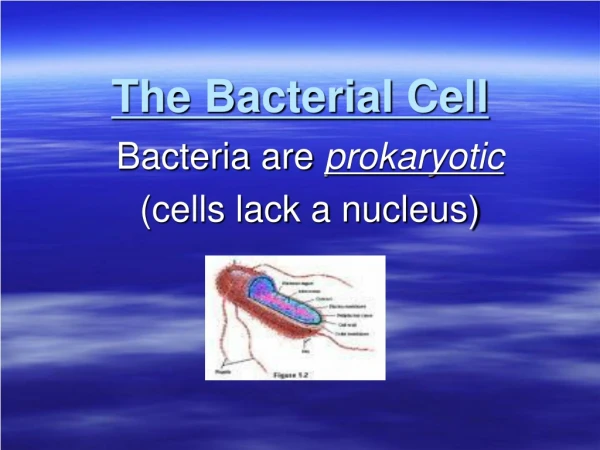
Bacterial Cell Structure. Prof.Dr . Rıza Durmaz YBÜ-2014. Object i ves. Discuss the basic parts and function of microscopes, Define the cell wall in prokaryotes Describe the functions of cell wall

Bacterial Cell Structure
E N D
Presentation Transcript
Bacterial Cell Structure Prof.Dr. Rıza Durmaz YBÜ-2014
Objectives • Discuss the basic parts and function of microscopes, • Define the cell wall in prokaryotes • Describe the functions of cell wall • Determine the differences of cell wall between gram positiveand gram negative bacteria and mycobacteria • Describe the functions of cytoplasmic membrane • Define the bacterial cytoplasm and internal structures : chromosome, plasmid, inclusion bodies, ribosome and endospore
OPTICAL METHODS • The light microscopes • Bright-field microscope • Dark-field microscope • Phase contrast microscope • Fluorescence microscope • Differential interference contrast microscope • The Electron microscope • Confocal scanning laser microscope • Scanning probe microscopes
Principles of Light Microscopy • They use visible light as a source of illumination • Thiermaximum magnification is 2000 times • Magnification- occurs in two phases • Objective lens- forms the real image • Ocular lens- forms the virtual image • Total power of magnification= the power of the objective X the power of the ocular • Theirresolutionpover is 0.2μm
Resolution • Resolution- the ability to distinguish two adjacent objects or points from one another • Also known as resolving power • Resolving power (RP) = Wavelength of light in nm 2 x Numerical aperture of objective lens • Under ideal conditions, resolving power of the light microscope is about half of the wavelength of the light being used. • With yellow light of a wavelength of 0.4 μm, the smallest separable diameters are about 0.2 μm • Shorter wavelengths provide a better resolution • Oil immersion lenses increase the numerical aperture
Bright-Field Microscopy • Most widely used in microbiology • These microscopes generally employ a 100-power objective lens with a 10-power ocular lens, • Magnifying the specimen 1000 times • Particles 0.2 μm in diameter are therefore magnified to about 0.2 mm and so become clearly visible. • The specimen produces an image that is darker than the surrounding illuminated field • These microscopes can be used with live, unstained and preserved, stain specimens
Dark-Field Microscopy • A bright-field microscope can be adapted to a dark-field microscope by adding a stop to the condenser • The stop blocks all light from entering the objective lens except for peripheral light • The specimen produces an image that is brightly illuminated against a dark field • It does not allow for visualization of fine internal details of cells • This technique has been particularly useful for observing organisms such as Treponemapallidum
Phase-Contrast Microscopy Transforms bright changes in light waves passing through a specimen into differences in light intensity Allows differentiation of internal components of live, unstained cells Useful for viewing intracellular structures such as bacterial spores, granules, and organelles
Interference Microscopy • Uses a differential-interference contrast (DIC) microscope • The image is colorful and three-dimensional • Allows for detailed view of live, unstained specimens, such as spores, vacuoles, granules
Fluorescence Microscopy Includes a UV radiation source and a filter that protects the viewer’s eyes Used with dyes that show fluorescence under UV rays Forms a colored image against a black field Used in diagnosing infections caused by specific bacteria, protozoans, and viruses using fluorescent antibodies
Confocal Microscopy Allows for viewing cells at higher magnifications using a laser beam of light to scan various depths in the specimen Most often used on fluorescently stained specimens Unstaining specimens can be visualized
Electron Microscopy • Magnification can be extremely high (between 5,000X and 1,000,000X for biological specimens) • Allows scientists to view the finest structure of cells • Viruses, cellwall, cytoplasm… • Two forms: transmission electron microscope (TEM) and scanning electron microscope (SEM)
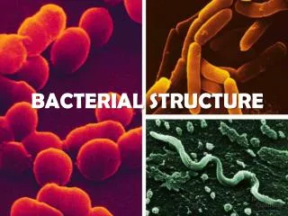
Bacteria can be distinguished from one another by their following characteristics: • morphology (size,shape,staining characteristics) • metabolic characteristics • Antigenic characteristics • Genetic characteristics
The size of bacterialcell • Perhaps the most obvious structural characteristic of bacteria is (with some exceptions) their small size. • For example, • Escherichia coli cells, an "average" sized bacterium, are about 2 micrometres (μm) long and 0.5 μm in diameter • Staphylococcus aureus: 1 μm in diameter • Bacillus anthracis: 1 μm in diameter and 3-4 μm in length • Brucellaabortus: 1.2μm in length • The average diameter of the Erythrocytes is ~7 µm • Granular leukocytes are ~ 12-15 µm in diameter
Bacterial Shapes ,ArrangementBacteria display a large diversity of cell morphologies and arrangements 1 Bacilli a. Bacillus (rod shape) b. Streptobacillus (chain formed bacillus e.g.Bacillussubtilis) c. Coccobacillus (very short and plump shaped bacillus e.g.Brucella) 2. Cocci a. Coccus (spherical shape) b. Diplococcus (two cells together e.g. Neisseria meningitidis) c. Streptococcus (chain formed coccus e.g. Streptococcus pyogenes) d. Staphylococcus (grape-like e.g. Staphylococcus aureus) e. Sarcina (packets of coccus) f. tetrads ( cocci in packets of four . e.g.Micrococcusspecies)
3. Spiral – helical, comma, twisted rod vibrio – gently curved or comma shaped spiral( Vibrio cholera) spirillum– thick, rigid, spiral; motility with flagella (Campylobacter, Helicobacter) spirochete– snake like thin,flexible spiral; motility with axial filament (Treponemapallidum, Borrelia)
Arrangement of cells is dependent on pattern of division and how cells remain attached after division.
Stains/dyes • Stains (dyes) are chemicals containing chromophores groups that impart color • Stains are generally salts in which one of the ions is colored • Based on the charges: • A basic dye consists of a colored cation (positively charged) with a colorless anion • Examples: crystal violet, safranin, basic fuchsin and methylene blue • Acid dyes have negatively charged chromophores. • They stain the background and leave the microbe transparent. Examples: Sodium eosinate, Nigrosine and congo red. • Neutral stain/dyes – stain with both charges
Based on function of stain • 1. Simple staining –Staining can be performed with basic dyes such as crystal violet or methylene blue • They are useful solely for increasing contrast so that morphology, size, and arrangement of organisms can be determined • 2. Differential staining - more than one dye is used- • Differentiation among bacteria is possible- • Eg. Gram’s staining, Acid-fast staining. • 3. Special staining – more than one dye used -Special structures are seen. • Eg. Capsule staining, Spore staining
Gram Stain • Gram staining allows clinicians to distinguish between two major classes of bacteria and to initiate therapy • Gram positive • Gram negative
Gram staining method • Fix the bacteria by heating or dry on to slide • Stain with crystal violet (purple color) • Precipitate the crystal violet with iodine • Wash with water and asetone based decoloriser to remove the unbound or excess stain • Add the safranin (red counter stain) to stain any decolorized cell • This process takes less than 10 min.
Principles of the Gram stain • Gram (+) bacteria turn purple, the stain gets trapped in peptidoglycan layer • Gram(-) bacteria have a thin peptidoglycan layer which does not retain crystal violet, cells counterstained with safranin and turned red.
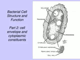
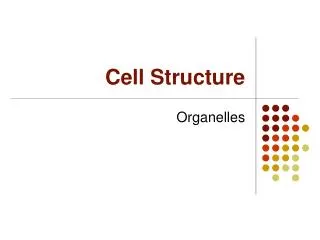
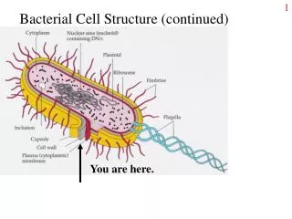
Bacterial Cell Structure • Appendages - flagella, pili or fimbriae • Surface layers - capsule, cell wall, cell membrane • Cytoplasm - nuclear material, ribosome, mesosome, inclusions etc. • Special structure - endospore
APPENDAGESFlagella: • a lash-like appendage • composed of helically coiled protein subunits(flagellin) • Some rods and spiral form bacteria have flagella • Bacteria may have one or several their surfaces function: a. Motility: Swimming toward food or away from poisons -chemical stimuli (chemotaxis) b.Antigenic: Carry antigenic and strain determinants origin : cell membrane, flagella attach to the cell by hook and basal body
onepolar flagella,monotrichous both ends, bipolar tuft at one end,lophotrishous all around bacteria,peritrichous
2. Fimbriae and Pili • Fimbriae: hairlike structures • They are composed of protein subunits (pilin) • Shorter, straighter, smaller than flagella • Not coiled in structure • Only on some gram negative (-) bacteria. • Several hundred are arranged peritrichously a) function: Adherence to other bacteria or to the host (alternative names adhesins,lectins,evasins,aggressins) • for the adherence of bacteria, specific receptor sites are required on the host cell membrane. • Not involve in motility. • Prevent phagocytosis • Fimbria is important virulence factor , Some pathogens cause diseases due to this Antigenic characteristic. e.g: E. coli, N. gonorrhoeae b) Origin: Cell membrane
pili • Sex pili (F pili): If bacteria have pili, they are F(+) or donors of F factor. • It is necessary for bacterial conjugation, to transfer DNA from one cell to another.
II. CELL SURFACE LAYER 1. Glycocalyx: Capsule and slime layer are also called glycocalyx. • It consists of polysaccharide or protein layers • Most of them have only polysaccharide. • B. anthracis has a capsule of poly-D-glutamic acid, • while S. pyogenes made of Hyaluronic acid • it is act as an barrier to toxic hydrophobic molecules (such as detergents) • it is a major virulans factor. Medically important (e.g.Streptococcuspneumoniae). • the capsule is antigenic
Capsule and Slime layer • Capsuleis well organized layer and not easily washed off • Slime layer, unorganized material, nonuniform and loosely adherent, that is easily removed. • They give mucoid growth on agar plate • Function: • Resistant to host phagocytosis, • Protect against desiccation, • Attachment to surface of solid objects. • They can be used for vaccine development • They can be used to serotyping of bacteria
Biofilm formation (surface bio-materials) Biofilm is a special bacterial adaptation that facilitates colonization • Sticky web of polysaccharides in glycocalyxbinds the cells together and to the surface • Biofilm is composed of numerous numbers of microorganisms • Biofilm is produced by certain bacteria (Coagulase negative staphylococci, S. aureus, P. aeruginosa, S.mutans ) • Biofilm forms at interfaces of Plastic surfaces orprosthetic devices • Within the biofilms, bacteria protect themselves from the immune system and from the effect of antibiotics (antibiotic resistance!) • Biofilms are important in human infections that are persistent and difficult to treat.
Axial Filaments • Theyare present in spirochetes ( Treponemapallidum causes syphilis) • Their function is motility – gliding motility • They look like as bundles of fibres at the ends of the cell • They are structurally similar to flagella • They locate under an outer membrane
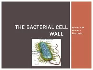
Bacterial Cell Wall • Most prokaryotes have peptidoglycan (murein=mucopeptide) layer. • The exceptions are • Archaeobacteria (which contain pseudoglycan) and • Mycoplasmas ( no cell walls) Peptidoglycan is unique to prokaryotic cells
Functions of the Cell Wall • Peptidoglycan provides rigidity, • it determines the shape of bacterial cell. • Contribute to pathogenicity (e.g. LPS in Gram negarivebacteria) • Protection from toxic compounds • Distinguish gram positive bacteria from gram negative bacteria • Protection for osmotic shock (lysis) • If water moves in lysis (in a hypotonic environment) • If water moves out plasmolysis (shriveling) (in a hypertonic environment)
Function of Cell wall-2 • The cell wall can be removed experimentally • Lysozyme – hydrolyzes PTG (peptidoglycan) • Penicillin – inhibits PTG syntesis • Result: • Gram-positive protoplasts • Gram-negative spheroplasts (OM intactandentrappedpeptidoglycan) • Bothformsare osmoticallysensitive • Theymust be maintained in an isotonic solution • L forms: Ifsuchcellsareabletogrowanddivide, theyarecalled L forms
L forms • L form: aredifficulttocultivateandrequirespecificagarmediumhavingtherightosmticstrength. • Some L form can reverttothe normal bacillary form. • Othersarestableandneverrevert. • SomebacterialspeciesproduceL formsspontaneously. • L formsmayproducechronicinfectionin host • L- form infectionsarerelativelyresistanttoantibiotictreatment, • Theirrevisiontothebacillary form can producerelapseof theovertinfection
Structure of Cell Wall • murein, mucopeptideorpeptidoglycan (allaresynonyms) • It is made up of linearpolysaccharidechainscross-linkedby peptides • Thebackbone of thepeptidoglycanconsist of N-acetylglucosamin (NAGA)andN-acetylmuramic acid (NAMA)connectedby 1-4 linkages.
CELL WALL cont… • TETRAPEPTIDES • Muramicasid (NAM) residuesarelinkedtoshortpeptides. • Thistetrapeptid is unusualbecause it containsboth L and D form aminoasids. • D-form a.a. is found in natureonly in prokaryoticcellwall. • Thecomposition of peptidesvariesonebacterialspeciestoanother.
Thetetrapeptidesidechainsof allspecieshavecertainimportantfeatures; • Mosthave L-alanine at position 1(attachedto NAM), D-glutamate at position 2, and D-alanine at position 4 • Poisition 3 is themostvariableone • Most gram-negativebacteriahavediaminopimelicacid at thisposition • Gram-positivebacteriausuallyhaveL-lysineat thisposition, howeversomemayhavediaminopimelicacidoranother amino acid. • Diaminopimelicacid is a unique element of bacterialcellwalls. • It is neverfound in thecellwalls of Archaeaoreukaryotes
Action of penicillin on cell wall synthesis Penicillin and other β-lactam antibiotics act by inhibiting penicillin-binding proteins, which normally catalyze cross-linking of bacterial cell walls. This is achieved through binding of penicillin to the enzymeDD-transpeptidase. As a consequence, DD-transpeptidase cannot catalyze formation of these cross-links
Gram positive bacterial cell wall • Thick cellwallis consist of multiplepeptidoglycanlayers(as many as 40 sheets in gram-positive, only 1 or 2 sheets in gram-negative) • In gram positive; pentaglycinbridgeis usedbetween tetrapeptide linkagestolengthenthecross-link( interpeptidbridges). In Gm (–) cellwall; interbridge is usedbetweentetrapeptide linkages
Gram positivecellwall Gram positivebacterialcellwall has teichoicacid Teichoicacid (TA):Polymer of riboseorglycerol joined by phosphate groups • Therearetwotypes of Teichoicasids: • Wall teichoicasidsarecovalentlylinkedtopeptidoglycan (WTA) • Lipoteichoicasidsarecovalentlylinkedtomembrane (LTA
Functions of teichoicacid • Constitutes major surface antigens of Gram positive species • Responsible for the negative charge of the cell surface • Participate in the supply of Mg++ to the cell by binding Mg++ • Regulate normal cell division • Resistance to environmental stresses, • such as heat , low osmolarity ,antimicrobial peptides, antimicrobial fatty acids, cationic antibiotics, and lytic enzymes produced by the host, including lysozymes • LTAs also act as receptors for phage particles • WTA act as a cementtostrengthenof peptidoglycan
Gram positive bacterial cell wall • Thepeptidoglycan can be degradedbytreatmentwithlysozyme(in serum,tissuesandsecretionsand in thephagocyticlysosome) • Withoutthepeptidoglycan, thebacterialyse. • Removal of cellwallwillproduces a protoplast. • Protoplastlysesunlessosmoticallystabilized.