The bacterial Cell Wall
The bacterial Cell Wall . Gram + & Gram – Bacteria. The Cell Wall. Is a complex , semi-rigid structure responsible for the shape of the cell as well as the size Surrounds the underlying, fragile plasma (cytoplasmic) membrane

The bacterial Cell Wall
E N D
Presentation Transcript
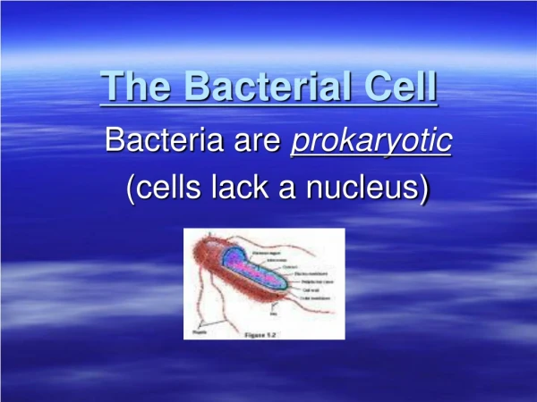
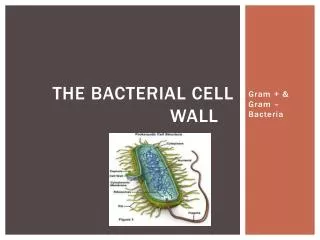
The bacterial Cell Wall Gram + & Gram – Bacteria
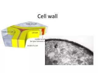
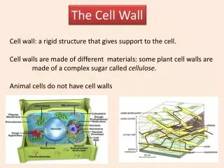
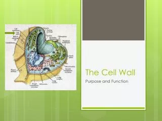
The Cell Wall • Is a complex, semi-rigid structure responsible for the shapeof the cell as well as the size • Surrounds the underlying, fragile plasma (cytoplasmic) membrane • Protects it and the interior of the cell from adverse changes in the outside environment • Major function is to prevent bacterial cells from rupturing • Osmotic lysis • Distinct Gram + and Gram - traits
Composition & Characteristics • Composed of macromolecular network called peptidoglycan • Peptidoglycanconsists of repeating disaccharideattached by polypeptides to form a lattice that surrounds and protects the entire cell • Disaccharideportion is made up of • Alternating rows of 10-65 sugars to form a carbohydrate “backbone” • Monosaccharides called N-acetylglucosamine (NAG) and N-acetylmuramicacid (NAM) • Adjacent rows are linked by polypeptides
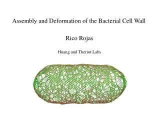
Peptidoglycan structure • Covalently attached to each NAM is a tetrapeptide chain • Tetrapeptide chains are linked by peptide cross-bridges • The result is a 3-D meshwork held together by covalent bonds Tetrapeptide chain Peptidoglycan Peptide bridge Tetrapeptide chain
Gram Positive (+) Cell Wall • Many layers of peptidoglycan • Thick layer (rigid structure) of peptidoglycan • Thicker than Gram – cell wall • Cell wall contains teichoic acids • Help in: • Attachment to surfaces • Provides rigidity • Helps in cell growth regulation • Two types • Lipoteichoic acid • Wall teichoic acid • Produce Exotoxins • Stains Purple during Gram Stain Lab test • Example: • Streptococcus pyogenes (strep throat)
Gram + Cell Wall What do the green spheres represent? What do the blue spheres represent?
Gram (+) and antibiotics • Analyze the cell wall of a Gram + bacteria • What part would be attacked by antibiotics and why? What would this do to the cell. Explain • http://faculty.ccbcmd.edu/courses/bio141/lecguide/unit1/prostruct/penres_fl.html
Gram Negative (-) Cell Wall • One or very few layers of peptidoglycan • Thin layer (not as thick as gram +) • Does NOT contain teichoic acids • Has an outer membrane outside the peptidoglycan layer • Consists of lipopolysaccharide (LPS), lipoproteins, phospholipids
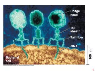
Gram (–) Cell wall • The outer membrane has several specialized functions • Its strong negative charge is an important factor in evading phagocytosis • Provides a barrier to certain antibiotics (for example penicillin), digestive enzymes, detergents • Permeability of outer membrane due to porins which allow passage of large molecules across the outer membrane • LPS (known as endotoxin) helps bacteria secrete toxins • Endotoxins and Exotoxins • Example: Escherichia coli (food poisoning) • StainsPinkin Gram Stain Lab test
Gram (-) and antibiotics • Analyze the Gram – bacterial structure • Why would Gram – bacteria be more resistant to antibiotics?
Gram Stain • Differences between Gram (+) and Gram (-) Bacteria: • Structural and functional differences between Gram-positive and Gram-negative cell walls can be used for identification and treatment of bacterial infections. • Basis for Gram stain (gram-positive = purple; gram-negative = pink)