Download
1 / 61
680 likes | 1.14k Vues
Cell Wall Inhibitors. Robert L. Copeland, Ph.D. 30 November 2014. Introduction.

E N D
Cell Wall Inhibitors Robert L. Copeland, Ph.D. 30 November 2014
Introduction The penicillins constitute one of the most important groups of antibiotics. Although numerous other antimicrobial agents have been produced since the first penicillin became available, these still are widely used, major antibiotics, and new derivatives of the basic penicillin nucleus still are being produced. Many of these have unique advantages, such that members of this group of antibiotics are presently the drugs of choice for a large number of infectious diseases.
History • The penicillins were the first antibiotics discovered as natural products from the mold Penicillium. In 1928, Sir Alexander Fleming, professor of bacteriology at St. Mary's Hospital in London, was culturing Staphylococcus aureus. He noticed zones of inhibition where mold spores were growing. He named the mold Penicillium rubrum. It was determined that a secretion of the mold was effective against Gram-positive bacteria.
1928 - Alexander Fleming • Bread mold (Penicillin notatum) growing on petri dish • 1939 - Florey, Chain, and Associates • Began work on isolating and synthizing large amounts of Penicillin. • 1944 - Used in WWII to treat infections • Late 1940’s - available for general use in US
Structure • Penicillins as well as cephalosporins are called beta-lactam antibiotics and are characterized by three fundamental structural requirements: • the fused beta-lactam structure (shown in the blue and red rings, • a free carboxyl acid group (shown in red bottom right), • one or more substituted amino acid side chains (shown in black). • The lactam structure can also be viewed as the covalent bonding of pieces of two amino acids - cysteine (blue) and valine (red).
thiazolidine ring (A) connected to a b-lactam ring (B), to which is attached a side chain (R).
The penicillin nucleus itself is the chief structural requirement for biological activity; • metabolic transformation or chemical alteration of this portion of the molecule causes loss of all significant antibacterial activity
Clinically useful families of beta-lactam compounds include the • penicillins, • cephalosporins, • monobactams • carbapenems
Classification • Penicillins (penicillin G) – greatest activity against gram+, gram-cocci, non-beta-lactamase-producing anaerobes • Antistaphylococcal penicillins (nafcillin) – resistant to staphylococcal beta-lactamases, active to staphylococci and streptococci • Extended-spectrum penicillins (ampicillin) – retain antibacterial spectrum of penicillin with improved activity against gram- organisms, but are destroyed by beta-lactamases.
Mechanisms of Drug Actions by Enzyme Inhibition: • All penicillin derivatives produce their bacteriocidal effects by inhibition of bacterial cell wall synthesis. Specifically, the cross linking of peptides on the mucosaccharide chains is prevented. If cell walls are improperly made cell walls allow water to flow into the cell causing it to burst.
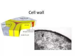
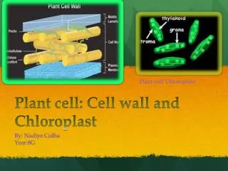
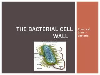
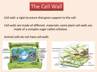
Cell Wall production • The cell walls of bacteria are essential for their normal growth and development. Peptidoglycan is a heteropolymeric component of the cell wall that provides rigid mechanical stability by virtue of its highly cross-linked latticework structure • The peptidoglycan is composed of glycan chains, which are linear strands of two alternating amino sugars (N-acetylglucosamine and N-acetylmuramic acid) that are cross-linked by peptide chains. (NAG-NAM). • In gram-positive microorganisms, the cell wall is 50 to 100 molecules thick, but it is only 1 or 2 molecules thick in gram-negative bacteria
The biosynthesis of the peptidoglycan involves about 30 bacterial enzymes and may be considered in three stages. • The first stage, precursor formation, takes place in the cytoplasm. The product, uridine diphosphate (UDP)-acetylmuramyl-pentapeptide, called a "Park nucleotide" accumulates in cells when subsequent synthetic stages are inhibited. • The last reaction in the synthesis of this compound is the addition of a dipeptide, D-alanyl-D-alanine. Synthesis of the dipeptide involves prior racemization of L-alanine and condensation catalyzed by D-alanyl-D-alanine synthetase.
The second stage, UDP-acetylmuramyl-pentapeptide and UDP-acetylglucosamine are linked (with the release of the uridine nucleotides) to form a long polymer. • The third and final stage involves the completion of the cross-link. This is accomplished by a transpeptidation reaction that occurs outside the cell membrane. The transpeptidase itself is membrane bound. The terminal glycine residue of the pentaglycine bridge is linked to the fourth residue of the pentapeptide (D-alanine), releasing the fifth residue (also D-alanine)
It is this last step in peptidoglycan synthesis that is inhibited by the beta-lactam antibiotics. • Penicillin binds at the active site of the transpeptidase enzyme that cross-links the peptidoglycan strands. It does this by mimicking the D-alanyl-D-alanine residues that would normally bind to this site. Penicillin irreversibly inhibits the enzyme transpeptidase by reacting with a serine residue in the transpeptidase. This reaction is irreversible and so the growth of the bacterial cell wall is inhibited.
Related targets for the actions of penicillins and cephalosporins; these are collectively termed penicillin-binding proteins (PBPs) • Covalently bind to a heterologous group of proteins called penicillin-binding proteins These PBPs may number 3 to 6 in any given bacteria. • There functions are diverse: catalyze the transpeptidase reaction, maintam shape, forms septums during division, Inhibit autolytic enzymes.
Binding to PBPs results in: • Inhibition of transpeptidase: transpeptidase catalyzes the cross-linking of the pentaglycine bridge with the fourth residue (D-Ala) of the pentapeptide. The fifth reside (also D-Ala) is released during this reaction. Spheroblasts are formed. • Structural irregularities: binding to PBPs may result in abnormal elongation, abnormal shape, cell wall defects.
Figure 45-2. The transpeptidase reaction in Staphylococcus aureus that is inhibited by penicillins and cephalosporins.
Figure 45-3. Comparison of the structure and composition of gram-positive and gram-negative cell walls.
Pharmacokinetics • Oral Administration of Penicillin G. About one-third of an orally administered dose of penicillin G is absorbed from the intestinal tract under favorable conditions. • Gastric juice at pH 2 rapidly destroys the antibiotic. The decrease in gastric acid production with aging accounts for better absorption of penicillin G from the gastrointestinal tract of older individuals. • Absorption is rapid, and maximal concentrations in blood are attained in 30 to 60 minutes. The peak value is approximately 0.5 unit/ml (0.3 mg/ml) after an oral dose of 400,000 units (about 250 mg) in an adult. • Ingestion of food may interfere with enteric absorption of all penicillins, perhaps by adsorption of the antibiotic onto food particles. Thus, oral penicillin G should be administered at least 30 minutes before a meal or 2 hours after. Despite the convenience of oral administration of penicillin G, this route should be used only in infections in which clinical experience has proven its efficacy.
Oral Administration of Penicillin V. The sole virtue of penicillin V in comparison with penicillin G is that it is more stable in an acidic medium, and therefore is better absorbed from the gastrointestinal tract. • On an equivalent oral-dose basis, penicillin V (K+ salt PEN-VEE K, V-CILLIN K, others) yields plasma concentrations two to five times greater than those provided by penicillin G. The peak concentration in the blood of an adult after an oral dose of 500 mg is nearly 3 mg/ml. Once absorbed, penicillin V is distributed in the body and excreted by the kidney in the same manner as penicillin G.
Parenteral Administration of Penicillin G • After intramuscular injection, peak concentrations in plasma are reached within 15 to 30 minutes. This value declines rapidly, since the half-life of penicillin G is 30 minutes. • Repository preparations of penicillin G are employed. The two such compounds currently favored are penicillin Gprocaine (maintained for as long as 4 to 5 days.) and penicillin G benzathine. (duration of antimicrobial activity in the plasma is about 26 day) • Such agents release penicillin G slowly from the area in which they are injected and produce relatively low but persistent concentrations of antibiotic in the blood. • Intrathecal administration is inadvisable particularly with benzylpenicillin as it can cause convulsions.
Distribution. • Penicillin G is distributed widely throughout the body, but the concentrations in various fluids and tissues differ widely. Its apparent volume of distribution is about 0.35 liters/kg. Approximately 60% of the penicillin G in plasma is reversibly bound to albumin. Significant amounts appear in liver, bile, kidney, semen, joint fluid, lymph, and intestine. • While probenecid markedly decreases the tubular secretion of the penicillins, this is not the only factor responsible for the elevated plasma concentrations of the antibiotic that follow its administration. Probenecid also produces a significant decrease in the apparent volume of distribution of the penicillins.
Cerebrospinal Fluid. Penicillin does not readily enter the CSF when the meninges are normal. However, when the meninges are acutely inflamed, penicillin penetrates into the CSF more easily. Although the concentrations attained vary and are unpredictable, they are usually in the range of 5% of the value in plasma and are therapeutically effective against susceptible microorganisms.
Excretion • Under normal conditions, penicillin G is rapidly eliminated from the body, mainly by the kidney but in small part in the bile and by other routes. Approximately 60% to 90% of an intramuscular dose of penicillin G in aqueous solution is eliminated in the urine, largely within the first hour after injection. • The half-time for elimination is about 30 minutes in normal adults (upto 10 hours in renal failure) . Approximately 10% of the drug is eliminated by glomerular filtration and 90% by tubular secretion. • Renal clearance approximates the total renal plasma flow. The maximal tubular secretory capacity for penicillin in the normal male adult is about 3 million units (1.8 g) per hour.
Clearance values are considerably lower in neonates and infants, because of incomplete development of renal function; as a result, after doses proportionate to surface area, the persistence of penicillin in the blood is several times as long in premature infants as in children and adults. • The half-life of the antibiotic in children less than 1 week old is 3 hours; by 14 days of age it is 1.4 hours. After renal function is fully established in young children, the rate of renal excretion of penicillin G is considerably more rapid than in adults.
Unitage of Penicillin • The international unit of penicillin is the specific penicillin activity contained in 0.6 mg of the crystalline sodium salt of penicillin G. One milligram of pure penicillin G sodium thus equals 1667 units; 1.0 mg of pure penicillin G potassium represents 1595 units. The dosage and the antibacterial potency of the semisynthetic penicillins are expressed in terms of weight. • The minimum inhibitory concentration(MIC) of any penicillin is usually given in ug/ml • Most penicillins ae dispensed as the sodium or potassium salt of the free acid.
Therapeutic Uses • Pneumococcal Infections • Pneumococcal Meningitis • Pneumococcal Pneumonia • Streptococcal Infections • Streptococcal Pharyngitis (including Scarlet Fever) • Streptococcal Pneumonia, Arthritis, Meningitis, and Endocarditis • Staphylococcal Infections • Meningococcal Infections • Gonococcal Infections • Syphilis • Actinomycosis • Diphtheria • Anthrax • Clostridial Infections • Fusospirochetal Infections • Rat-Bite Fever • Listeria Infections • Lyme Disease • Erysipeloid • Surgical Procedures in Patients with Valvular Heart Disease
The antimicrobial activity of carbenicillin, its indanyl ester (carbenicillin indanyl), and ticarcillin is extended to include Pseudomonas, Enterobacter, and Proteus species. • Other extended-spectrum penicillins include mezlocillin and piperacillin, which have useful antimicrobial activity against Pseudomonas, Klebsiella, and certain other gram-negative microorganisms.
Mechanisms of Bacterial Resistance to Penicillins • Resistance to penicillins and other beta lactams is due to one of four general mechanisms: • Inactivation of the antibiotic by beta lactamase • Modification of target PBPs • Imparied penetration of drug to target PBPs • The presence of an efflux pump.
There are more than 300 different types of this enzyme. The process is genetically controlled commonly with plasmids. • beta-lactamase production is particularly important in Staphylococci but other organisms such as Neisseria gonorrhoeae and Hemophilus species also produce these enzymes where as beta-hemolytic Streptococci do not. • • In developed countries at least 80% of Staphylococci now produce beta-lactamase.
Other resistance mechanisms • A reductionin the permeability of the outer membrane. • Thus there is a decreased ability of the drug to penetrate to the target site. • The occurrence of modified penicillin binding sites. This mechanism is responsible in methicillin resistance in Pneumococci.
The penicillins described in this section are resistant to hydrolysis by staphylococcal penicillinase. Their appropriate use should be restricted to the treatment of infections that are known or suspected to be caused by staphylococci that elaborate the enzyme¾the vast majority of strains of this bacterium that are encountered in the hospital or in the general community. These drugs are less active than is penicillin G against other penicillin-sensitive microorganisms, including non-penicillinase-producing staphylococci. • The penicillinase-resistant penicillins remain the agents of choice for most staphylococcal disease, despite the increasing incidence of isolates of so-called methicillin-resistant microorganisms.
Adverse effects • Hypersensitivity Reactions. Hypersensitivity reactions are by far the most common adverse effects noted with the penicillins, and these agents probably are the most common cause of drug allergy. There is no convincing evidence that any single penicillin differs from the group in its potential for causing true allergic reactions. • The basis of which is the fact that degradation products of penicillin combine with host protein and become antigenic.
These are cross-reactions between various types of penicillins. • In approximate order of decreasing frequency, manifestations of allergy to penicillins include maculopapular rash, urticarial rash, fever, bronchospasm, vasculitis, serum sickness, exfoliative dermatitis, Stevens-Johnson syndrome, and anaphylaxis The overall incidence of such reactions to the penicillins varies from 0.7% to 10% in different studies. • Very high doses of penicillin G can cause seizures in kidney failure.
Stevens Johnson Syndrome Adverse drug reactions. • Painful Blistering of the skin and mucous membrane involvment. • In many cases preceded with flu like symptoms and high fever. • As it evolves the skin literally sloughs off. • Ocular involvement includes severe conjunctivis, iritis, palpebral edema, conjunctival and corneal blisters and erosions, and corneal perforation.
Management of the Patient Potentially Allergic to Penicillin. • Evaluation of the patient's history is the most practical way to avoid the use of penicillin in patients who are at the greatest risk of adverse reaction. The majority of patients who give a history of allergy to penicillin should be treated with a different type of antibiotic. • "Desensitization" occasionally is recommended for patients who are allergic to penicillin and who must receive the drug. This procedure consists of administering gradually increasing doses of penicillin in the hope of avoiding a severe reaction and should be performed only in an intensive care setting.
Apparent toxic effects that have been reported include bone marrow depression, granulocytopenia, and hepatitis. The last-named effect is rare but is most commonly seen following the administration of oxacillin and nafcillin. • Regardless of the route by which the drug is administered, but most strikingly when it is given by mouth, penicillin changes the composition of the microflora by eliminating sensitive microorganisms. This phenomenon is usually of no clinical significance, and the normal microflora is reestablished shortly after therapy is stopped. In some persons, however, superinfection results from the changes in flora. Pseudo-membranous colitis, related to overgrowth and production of a toxin by C. difficile, has followed oral and, less commonly, parenteral administration of penicillins.
Adverse effects cont • Convulsions and encephalopathy can occur, especially at higher doses and especially if administered intrathecally (NOT advised). • Interstitial nephritis (Methicillin) • Coomb's positive hemolytic anemia • Neutropenia (especially the b-lactamase -resistant penicillins) • Decreased platelet aggregation (carbenicillin and ticarcillin) • Hypernatremia and hypokalemia (carbenicillin)
Drug-drug Interactions • Penicillins bind to and inactivate aminoglycosides. This is a form of chemical antagonism. If an aminoglycoside and a penicillin are combined. they MUST NOT be administered simultaneously through the same I.V. line or through the same syringe. They will crystallize and precipitate in the line or in the vessels! • When an aminoglycoside and a penicillin are administered, the infusions should be staggered by about 1 to 2 hours.
Classification of the Penicillins and Summary of Their Pharmacological Properties • 1. Penicillin G and its close congener penicillin V are highly active against sensitive strains of gram-positive cocci, but they are readily hydrolyzed by penicillinase. Thus, they are ineffective against most strains of Staphylococcus aureus. • 2. The penicillinase-resistant penicillins (methicillin, nafcillin, oxacillin, cloxacillin, and dicloxacillin) have less potent antimicrobial activity against microorganisms that are sensitive to penicillin G, but they are effective against penicillinase-producing Staph. aureus. • 3. Ampicillin, amoxicillin, bacampicillin, and others comprise a group of penicillins whose antimicrobial activity is extended to include such gram-negative microorganisms as Haemophilus influenzae, E. coli, and Proteus mirabilis. Unfortunately, these drugs and the others listed below are hydrolyzed readily by broad-spectrum b-lactamases that are found with increasing frequency in clinical isolates of these gram-negative bacteria.
Cephalosporins • Mechanism of Action: Cephalosporins are composed of a dihydrothiazine ring and a b-lactam ring. The mechanism of action is identical to penicillins. • Mechanism of Resistance: Same as penicillins. • Cephalosporins are less susceptible to Staphylococcus beta-lactamase; therefore have a broader spectrum of activity; however they are not the drug of choice. Other bacteria are resistant, because they produce distinct beta-lactamases. Methicillin-resistant Staphylococcus is resistant to most cephalosporins. • Classification: The cephalosporins are classified as first, second, third generation or forth generation cephalosporins. This classification is dependent on the antimicrobial activity.
Penicillins Cephalosporins
Cephalosporins First Generation Second Generation Third Generation Fourth Generation * Oral Agent
First generation cephalosporins: • cephalothin, cefazolin, cefalexin. These drugs have good activity against most Gram positive cocci (Streptococcus, pneumococcus but not or methicillin-resistant Staphylococcus). They are more active against Gram negative organisms (Escherichia co1i Kiebsiella pneumoniae, and the indole negative Proteus mirabilis) than are the natural penicillins. They are effective against some anaerobic cocci (Peptococcus and Peptosteptococcus, but NOT Bacteroides fragilis). • They are ineffective against Pseudomonas aeruginosa, Enterobacter, and indole-positive Proteus species. • These drugs do not cross the blood-brain barrier.
Second generation cephalosporins: • cefuroxime, cefamandole, cefoxitin, cefaclor. The spectrum is extended to more Gram negative bacteria Enterobacter species, Klebsiella species, and indole-positive Proteus species. Also, Haemophilus influenza is covered by cefuroxime, cefamandole, cefaclor; Bacteroides fragilis by cefoxitin. • These drugs do not achieve adequate levels in the CSF.
Third generation cephalosporins: • moxalactam, cefaperazone, ceftazidirne, ceftriaxone. These drugs demonstrate extended Gram negative coverage, are more resistant to non-Staphylococcus b-lactamase, and readily cross the blood-brain barrier. The spectrum is extended to include: Enterobacter, Pseudomonas (ceftazidime and cefaperazone only), Serratia, b-lactamase producing Haemophillus influenza and Neisseria species. • Only cetizoxime and moxalactam retain good activity against Bacteroides fragilis.
Fourth generation • forth generation of cephalosporins (e.g. cefepime) are available, these are comparable to third-generation but more resistant to some betalactamases.
Pharmacokinetics • Some cephalosporins may be given orally but most are given parenterally (IM or IV). • They are widely distributed in the body like penicillins. • Some such as cefoperazone, cefotaxime, cefuroxime, ceftriaxone, and ceftazidime (third generation) also cross the blood-brain barrier and are drugs of choice for meningitis due to Gram-negative intestinal bacteria.