
RESPIRATORY SYSTEM
RESPIRATORY SYSTEM. Our chosen topic is: PLEURAL EFFUSIONS DEFINITION - A COLLECTION OF FLUID BETWEEN THE PARIETAL PLEURA AND VISCERAL PLEURA . SYMPTOMS - Chest pains - Shortness of Breathe -Cough -Hiccups -Fever.

RESPIRATORY SYSTEM
E N D
Presentation Transcript
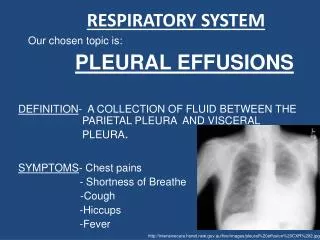
RESPIRATORY SYSTEM Our chosen topic is: PLEURAL EFFUSIONS DEFINITION- A COLLECTION OF FLUID BETWEEN THE PARIETAL PLEURA AND VISCERAL PLEURA. SYMPTOMS- Chest pains - Shortness of Breathe -Cough -Hiccups -Fever http://intensivecare.hsnet.nsw.gov.au/five/images/pleural%20effusion%20CXR%202.jpg
ANATOMY AND PHYSIOLOGY IN A HEALTHY LUNG The Right Lung -Makes up 56% of the total lung volume -Three lobes-the superior, middle and inferior, which are separated by the horizontal fissure and the oblique fissure. http://home.comcast.net/~wnor/thoraxlesson2.htm The Left Lung -Makes up 44% of the total lung volume -Two lobes which are separated by the oblique fissure. http://home.comcast.net/~wnor/thoraxlesson2.htm
The main anatomy affected by pleural effusions are the layers in the Lung- • There are two layers-the parietal pleura and the visceral pleura. • At the Hilum, the parietal pleura folds back on itself to become the visceral pleura. • The mesothelium is a membrane that forms the lining of the • pleura. It includes the pleura, the pericardium and the • peritoneum. • Function- to produce a lubricating fluid that is secreted between • the parietal pleura and the visceral pleura to reduce friction and • provide a non-adhesive, protective surface. • The volume of pleural fluid is around 10ml in a healthy lung and has a pH of 7.60-7.64 • The pleural fluid contains – • glucose content similar to that of plasma • mesothelial cells • macrophages • lymphocytes • sodium, potassium and calcium concentrations similar to that of interstitial fluid. • Lactate Dehydrogenase concentration of less than 50% of that of plasma http://www.nature.com/modpathol/journal/v18/n2/images/3800278f1.jpg
ANATOMY OF A HEALTHY LUNG ANATOMY OF A LUNG WITH A PLEURAL EFFUSION http://www.themesotheliomalibrary.com/pleural-effusions3.gif A pleural effusion is an accumulation of fluid between the parietal pleura and the visceral pleura.
ANATOMY OF A LUNG WITH A PLEURAL EFFUSION • The fluid tends to be pleural fluid. • The fluid accumulates due to the over production of pleural fluid by the mesothelial cells and separates the visceral and parietal pleura. • This fluid can not be drained by the lymphatic system, and so • therefore continues to accumulate, resulting in a pleural effusion. • The accumulation of fluid may also be due to changes in • hydrostatic pressure or oncotic pressure. • The contents and pH of the pleural fluid alters, depending • on whether the effusion is Transudative or Exudative • Other changes to anatomy include- • -Pleural thickening • -Mediastinal Pleural thickening • -Diaphragmatic Pleural thickening • -Presence of Pleural nodules which can range from 2mm to 4cm • -Increase in pressure, due to the increase in fluid www.ncbi.nlm.nih.gov


