Download
1 / 19
190 likes | 219 Vues
This article provides a critical evaluation of the published literature on the structural basis for Duffy recognition and erythrocyte invasion by the malaria parasite. It analyzes the structural characteristics of the Duffy-binding-like (DBL) domain and its role in invasion and cytoadherence. The study highlights the different binding sites and substrates targeted by various DBL domains and suggests potential therapeutic strategies.

E N D
Critical Evaluation of Published Literature BMSD 542, Winter 2006 Structural basis for Duffy recognition by the malaria parasite Duffy-binding–like domain Singh SK, Hora R, Belrhali H, Chitnis CE, Sharma A Nature 439: 741-744, 2005 Structural basis for the EBA-175 erythrocyte invasion pathway of themalaria parasite Plasmodium falciparum Tolia NH, Enemark EJ, Sim KL, Joshua-Tor L Cell 122: 183-193, 2005
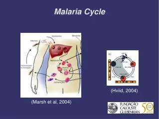
Background Plasmodium causes Malaria which affects more than 500 million people and kills about two million annually. P. falciparum is the most prevalent; P. vivax less so; P. knowlesi is the simian counterpart. Infection is caused by sporozoites entering into the host bloodstream after a female Anopheles mosquito bite and infecting hepatocytes; the hepatocytes rupture and release thousands of merozoites each of which can invade an erythrocyte, thus initiating the asexual erythrocytic stage of the parasite’s life cycle. All pathological and clinical manifestations of the disease are caused by this critical invasion step.
Background, continued… Binding to the host endothelium is accomplished through the function of a common adhesion molecule found in two families of parasite ligands. EBL (erythrocyte binding ligand) family of erythrocyte invasion ligands and the var/PfEMP1 (P. falciparum erythrocyte membrane protein 1) family of cytoadherence ligands This adhesive domain, called the Duffy-binding like (DBL) domain was first described as part of the Duffy Binding Protein (DBP), an important invasion ligand of both P. vivax and P. knowlesi for erythrocytes. DBL domains contribute to both invasion and cytoadherence and are one of the most versatile and polymorphic adhesive molecules. X-ray crystallography structures of the DBL domains from two well-studied EBL ligands, the P. knowlesi DBP (PkDBP) and the P. falciparum EBA-175, have been published.
EBP, EBA, PfEMP, … Nature 439: 741-744, 2005
EBL, DBL, DBP, PfEMP, … erythrocyte binding domain (critical) Cell 122: 183-193, 2005
PkDBP alignments Nature 439: 741-744, 2005
EBA-175 alignments Cell 122: 183-193, 2005
PkDBP structure (3 Å) 11 alpha helices, unique fold, three subdomains Nature 439: 741-744, 2005
EBA-175 structure (2.3 Å) Cell 122: 183-193, 2005
Binding DBL domains bind several substrates and have different binding sites, sometimes involving dimerisation. EBA-175 and PkDBP bind different receptors on the erythrocyte surface. PkDBL (and PvDBL) bind the host DARC (Duffy antigen receptor for chemokines); mutagenesis data available. EBA-175, BAEBL, and JSEBL all can bind sialic acid residues, but each recognises different erythrocyte sialoglycoproteins; receptor for EBA-175 is human RBC receptor glycophorin A. Redundant pathways means that EBA-175 is not essential. X-ray structure of EBA-175 was solved with sialic acid derivative, alpha-2,3,sialyllactoase to identify the glycan binding site; additional mutagenesis experiments were performed.
PkDBL binding Nature 439: 741-744, 2005
PvDBL binding polymorphic residues in field isolates; immune system evasion binding site “just-in-time” release and binding to receptor Nature 439: 741-744, 2005
PkDBL binding SD2 DARC binding site antigenic site SD2 SD1 antigenic site
EBA-175 binding Cell 122: 183-193, 2005
Model for EBA-175 region 2 binding to glycophorin A Cell 122: 183-193, 2005
Superimposed DBLs (within 2 Å of each other) DARC binding site glycan binding sites
A treasure of insights DARC binding site glycan binding site
Conclusions – insights from x-ray structures Different sites target different substrates and receptors in different DBL domains Interplay between sequence conservation and variation to evade immune evasion For PvDBL, target the DBL-DARC interaction for therapeutics (conserved) For EBA-175, target the glycan binding sites; distrupt dimersation and/or DBL domain interactions