Download
1 / 58
600 likes | 729 Vues
Discover the interdependent systems of shoot and root in plants, from tissue systems to meristems. Learn about the functions, morphology, and types of plant cells in detail.

E N D
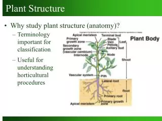
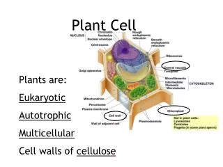
Shoot and Root Systems • Shoot system • produces sugars by photosynthesis • carries out reproduction Shoot System • Root system • anchors the plant • penetrates the soil and absorbs water and minerals • stores food Root System
Shoot and root systems are interdependent water & minerals sugar SHOOT SYSTEM ROOT SYSTEM
Plant Tissue Systems EPIDERMIS • Ground tissue system • Vascular tissue system • Dermal tissue system VASCULAR TISSUES GROUND TISSUES SHOOT SYSTEM ROOT SYSTEM
Meristems – Where Cells For New Organs Originate • Regions where cell divisions produce plant growth • Apical meristems • Lengthen stems and roots • Responsible for primary growth • Lateral meristems • Increase width of stems • Responsible for secondary growth
Apical Meristems Lengthen shoots and roots activity at meristems new cells elongate and start to differentiate into primary tissues Cells that form at apical meristems: protoderm epidermis ground meristem ground tissues procambium primary vascular tissues
Lateral Meristems Increases girth of older roots and stems Cylindrical arrays of cells vascular cambium secondary vascular tissues periderm cork cambium thickening
Figure 35.7 The three tissue systems The Three Tissue Systems in Plants
Simple Tissues Made up of only one type of cell Parenchyma Collenchyma Sclerenchyma
Morphology of Three Simple Tissue Types parenchyma collenchyma sclerenchyma
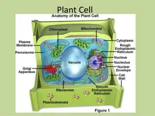
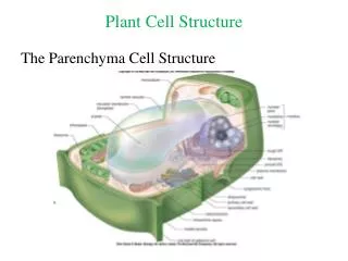
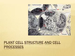
Parenchyma: A Simple Tissue • Comprises most of a plant’s soft primary growth • Cells are pliable, thin walled, many sided • Cells remain alive at maturity and retain capacity to divide • Mesophyll is a type of parenchyma that contains chloroplasts
Collenchyma: A Simple Tissue • Specialized for support for primary tissues • Cells are elongated, with walls (especially corners) thickened with pectin • Makes stems strong but pliable • Cells are alive at maturity
Sclerenchyma: A Simple Tissue • Supports mature plant parts • Protects many seeds • Cells have thick, lignified walls and are dead at maturity • Two types: • Fibers: Long, tapered • cells • Sclereids: Stubbier cells
Complex Tissues Composed of a mix of cell types Xylem Phloem Epidermis
Xylem • Conducts water and dissolved minerals • Conducting cells are dead and hollow at maturity vessel member tracheids
Phloem: A Complex Vascular Tissue • Transports sugars • Main conducting cells are sieve-tube members • Companion cells assist in the loading of sugars sieve plate sieve-tube member companion cell
Epidermis: A Complex Plant Tissue - Covers and protects plant surfaces - Secretes a waxy, waterproof cuticle • In plants with secondary growth,periderm replaces epidermis
Monocots and Dicots – same tissues, different features 1 cotyledon 2 cotyledons 4 or 5 floral parts 3 floral parts Netlike veins Parallel veins 3 pores 1 pore Vascular bundles dispersed Vascular bundles in ring
Stems – • organs consisting of an alternating • system of nodes, the points at which • leaves are attached, and internodes, • the stem segments between nodes. • the main functions of the stems include • conducting sugars and water and holding • leaves up into the sunlight
Monocot and Dicot Stems (Two Divisions of Angiosperms) Dicot Stem Monocot Stem
Bud = undeveloped shoot of meristematic tissue Leaves Internode spaces between leaf attachments Axillary bud at node (can form lateral shoots) Longitudinal section of terminal bud
Shoot Development shoot apical meristem procambrium ground meristem protoderm cortex procambrium pith primary xylem primary phloem
Internal Structure of a Dicot Stem - Outermost layer is epidermis - Cortex lies beneath epidermis - Ring of vascular bundles separates the cortex from the pith - The pith lies in the center of the stem
Internal Structure of a Monocot Stem • The vascularbundles arescattered throughout the ground tissue • No division of ground tissue into cortex and pith
Secondary Growth • Occurs in perennials • A ring of vascular cambium produces secondary xylem and phloem • Wood is the accumulation of these secondary tissues, especially xylem
Woody Stem periderm (consists of cork, cork cambium, and secondary cortex) secondary phloem SAPWOOD HEARTWOOD BARK vascular cambium
Annual Rings • Concentric rings of secondary xylem • Alternating bands of early and late wood • Early wood • Xylem cells with large diameter, thin walls • Late wood • Xylem cells with smaller diameter, thicker walls
Types of Wood • Hardwood (oak, hickory) • Dicot wood • Xylem composed of vessels, tracheids, and fibers • Softwood (pine, redwood) • Gymnosperm wood • Xylem composed mostly of tracheids • Grows more quickly
Adapted for Photosynthesis • Leaves are usually thin • High surface area-to-volume ratio • Promotes diffusion of carbon dioxide in, oxygen out • Leaves are arranged to capture sunlight • Are held perpendicular to rays of sun • Arrange so they don’t shade one another
Leaf Structure UPPER EPIDERMIS cuticle PALISADE MESOPHYLL xylem SPONGY MESOPHYLL phloem LOWER EPIDERMIS Stoma with guard cells CO2 one stoma O2
Mesophyll:Photosynthetic Tissue • A type of parenchyma tissue • Cells have chloroplasts • Two layers in dicots • Palisade mesophyll • Spongy mesophyll
Collenchyma Parenchyma
Leaf Veins: Vascular Bundles • Xylem and phloem – often strengthened with fibers • In dicots, veins are netlike • In monocots, they are parallel
Root Structure • Root cap covers tip • Apical meristem produces the cap • Cell divisions at the apical meristem cause the root to lengthen • Farther up, cells differentiate and mature root apical meristem root cap
Fibrous Tap Root Systems Lateral Roots grow from the Tap Root
epidermis endodermis cortex pericycle root hair phloem xylem
cortex epidermis endodermis pericycle xylem phloem Cross Section of a Root
Internal Structure of a Root • Outermost layer is epidermis • Root cortex is beneath the epidermis • Endodermis, then pericycle surround the vascular cylinder • In some plants, there is a central pith
Root Hairs and Lateral Roots • Both increase the surface area of a root system • Root hairs are tiny extensions of epidermal cells • Lateral roots arise from the pericycle and must push through the cortex and epidermis to reach the soil new lateral root
Lateral Root Figure 35.16 The formation of lateral roots
Figure 36.7 Lateral transport of minerals and water in roots