Download
1 / 77
820 likes | 945 Vues
Learn about various techniques for isolating enzymes and purifying them, including extraction, fractionation, and purification methods. Understand the importance of enzyme source selection and quality control in industrial applications.

E N D
A. Pendahuluan • Beberapa teknik isolasi enzim secara umum dikelompokan menjadi: • Metode klasik berupa distilasi dan ekstraksi dengan pelarut organik (berdasarkan sifat umum makromolekul: pH, kekuatan ion dan kelarutan) Kurang digunakan lagi karena kaitannya dengan kestabilan enzim • Perbedaan sifat protein globular: Mr, muatan protein • Interaksi spesifik dan reversibel antara enzim dengan substrat, koenzim, ligan
PEMISAHAN MATERIAL • Pengambilan bahan tidak larut (Removal of Insolubles). Sedikit mengkonsentrasikan produk atau perbaikan produk. Filtrasi dan sentrifugasi. • Isolasi Produk. Tidak spesifik, pengambilan bahan yang mempunyai sifat yang tersebar dibandingkan dengan produk yang diinginkan. Konsentrasi dan kwalitas produk mulai terjadi. Adsorpsi dan ekstraksi solven. • Purifikasi. Teknik proses yang sangat selektif untuk menghasilkan produk dan mengambil bahan yang tidak diinginkan serupa dengan fungsi kimia dan sifat fisika. Khromatografi, elektrophoresis, dan presipitasi. • Produk akhir. Kristalisasi
B. Tahapan isolasi • Lokasi enzim: • Ekstraseluler (ekoenzim) • Endoseluler (terikat pada partikel subseluler atau membran sel) • Hal yang perlu diperhatikan • Sifat khas materi utama • Bentuk (cair, padat) • Tipe umum (animal, vegetal, mikroba) • Struktur biologi (seluler, tisuler) • Lokasi produk yang dicari: mitokondria, sitoplasma, membran, dll.
Sifat struktural dan fisiko-kimia: • Mr, struktur molekul, stabilitas • pH optimum, pI • Aktivator, inhibitor • Konstanta kinetika • Pelepasan protein dalam bentuk cair tanpa menghilangkan aktivitas • Tanpa merusak struktur • Temperatur rendah (4ºc) • Penggunaan buffer • Penggunaan reagen pelindung (EDTA, 2-mercaptoethanol, substrat, dll • Perlakuan secara cepat dan hati-hati
B.1. Tahapan Isolasi • Materi utama sangat heterogen, maka untuk mendapatkan enzim murni perlu tahapan isolasi • Ekstraksi: peelepasan enzim dari sel atau bagian sel dan didapatkan ekstrak dalam bentuk cair yang mempunyai sifat fisiko kimia sama • Fraksionasi: memisahkan ekstraks berdasarkan kelarutannya guna mendapatkan kelompok molekul yang sama (fraksi) • Purifikasi: pemisahan fraksi lebih lanjut dengan metode fisiko-kimia atau biospesifik untuk mendapatkan molekul enzim lebih murni
Skema umum isolasi dan purifikasi enzim Materi Primer Animal Mikroba Vegetal Tahap Ekstraksi Ekstrak total Tahap Fraksionasi Ekstrak kasar Tahap Purifikasi Enzim murni
Microbial source • Often more stable than analogous enzyme obtained from plant or animal tissue • Generally Recognized As Safe (GRAS) certified microbes are non pathogenic, nontoxic, and generally they do not produce antibiotics
Plant source • Represent a traditional source of a wide range of enzymes • Plant tissues are chosen as a source for subsequent purification of various enzymes
Animal source • Animal tissues are a source of several enzymes of industrial use and therapeutic use • The other organs like stomach, placenta, heart, kidney or cells like erythrocytes can be sources for specific enzymes
STEP IN ENZYME PURIFICATION Enzyme extract Crude Enzyme Dilute Enzyme Conc. Enzyme
Teknik separasi dan purifikasi berdasarkan sifatnya Ultrasentrifugasi Dialisa Gel Filtrasi Kromatografi penukar ion Elektroforesis Ultrasentrifugasi Sentrifugasi zonal Mobilitas elektroforesis Gel Elektroforesis Mr Muatan Elektro dekantasi Enzim Densitas Titik isoelektrik Kelarutan Sifat permukaan Stabilitas Pengendapan Isoelektrik Kromatografi Adsorpsi • Perlakuan : • asam-basa • suhu • Kromatografi: • Afinitas • Hidrofobik • Kovalen Partisi Cair-cair Pengendapan terfraksi dengan garam atau pelarut organik
B.2. Kontrol Kualitas • Enzim industrial perlu adanya kontrol kualitas yang meliputi; • Kemurnian enzim dalam periode waktu tertentu (aktivitas spesifik) • Kontrol kemurnian dengan metode fisiko-kimia; homogenitas dan sifat karakteristiknya (Mr, polimorfisme...) • Uji stabilitas: resiko denaturasi, semakin murni enzim semakin mudah terdenaturasi. Perlu dilakukan: • Eliminasi kontaminan • Penyimpanan pada temperatur renadah • pH netral
Prosedur umum kontrol kwalitas enzim murni • Pengukuran : • Aktivitas katalitik • Protein • Aktivitas spesifik • Kontaminan (enzim & lainnya • Elektroforesis • Ultrasentrifugasi • Gel Filtrasi • Pengukuran: • Tekanan osmose • Pengendapan dengan UF • Difusi dan koefisien difusi • Gel eksklusi kromatografi Aktivitas biologi & Spesifik Homogenitas SDS PAGE Mr Enzim murni Metode Sanger Polimorfisme, Mr Komposisi & Sequence Kemasan Stabilitas Label Penggunaan
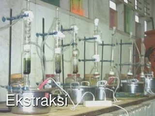
C. Ekstraksi • Ekstraksi; pelepasan enzim dari sel atau bagian sel menggunakan proses mekanik, dan non mekanik (kimia, enzimatis, dll) • Jaringan vegetal dan animal: penghalusan dan homogenisasi, secara mekanik • Sel mikroorganisme secara umum adalah pemecahan dinding sel secara mekanik dan non mekanik • Kimia: alkali/asam, deterjen, osmose, EDTA memecah bakteri gram negatif • Enzimatis: lisosim (memutus 1-4 glukosida peptidoglican)
Pemecahan dinding mikroorganisme • Proses mekanik: • Ultrasonikasi: sel dipecah • Pembekuan-pencairan • Penggerusan/agitasi dengan partikel gelas • Desintegrasi pada P> • Proses non-mekanik: • Desikasi dengan spray drying • Liase secara kimia dan fisika • Perlakuan alkali • Deterjen: Na-lauril sulfat, trixtron X-100 • Shock dingin : jumlah kecil • Shock osmotic: perubahan konsentrasi garam • Liase Enzimatik • Lisozim : hidrolisis beta 1,4 glukosida • Autolisis: dengan proteolise, lipolise
D. FRAKSIONASI D.1. FRAKSIONASI PENGENDAPAN D.1.1. PENGENDAPAN DENGAN GARAM • Garam yang paling banyak digunakan Amm. Sulfat • Prinsip: • Protein larut dalam larutan garam pada pH sekitar pI • Kelarutan lebih kuat dibanding dengan kekuatan ion dalam larutan (salting in) • Batas kekuatan ion tertentu, kelarutan berkurang (salting out) berkaitan dengan terhidrasinya protein
Daerah pengendapan bergantung pada: • Garam yang digunakan • Jenis proteinnya • Kelebihan (NH4)2SO4: • Harganya murah • Kemampuan pengendapan tinggi • Kelarutannya besar, endotermik • Efek denaturasi terhadap protein rendah • Kristalisasi garam tertentu yang terendapkan oleh konsentrasi garam tertentu dapat dilarutkan kembali dengan melarutkannya pada pelarut dengan kadar lebih rendah
pI Kelarutan minimum, mengendap D.1.2. PENGENDAPAN PADA ISOELEKTRIK D.1.3. EFEK TEMPERATUR Pengaturan pH larutan Protein Globular Hemoglobin T>Kelarutan>, s/d 40-50C Pengaturan T Seleksi Protein Protein terdenaturasi Tidak dapat digunakan secara industri
D.1.2. PENGENDAPAN DENGAN PELARUT ORGANIK • Alkohol • Isopropanol (paling banyak digunakan) untuk enzim ekstraseluler; amiloglukosidase • Methanol • Aseton dan etil eter: protein sedikit larut maka perlu jumlah yang banyak Protein Saling bergabung Protein mengendap Enzim <: konstanta dielektrikum & kestabilan Penambahan pelarut pada T< shg tidak mendenaturasi Tambah pelarut organik
Electrophoresis • Principle is to separate proteins (in tact) on the basis of their charge and their ability to migrate within a gel (jello-like) matrix • A strong electric field is applied to the protein mixture for an extended period of time (hours) until the proteins move apart or migrate
Isoelectric Point (pI) • The pH at which a protein has a net charge=0 • Q = S Ni/(1 + 10pH-pKi) Transcendental equation
Increasing pH _ _ _ _ _ _ _ _ _ + + + + + + + + + C A T H O D E A N O D E pI = 8.6 pI = 6.4 pI = 5.1 IEF Principles
Isoelectric Focusing • Separation of basis of pI, not Mw • Requires very high voltages (5000V) • Requires a long period of time (10h) • Presence of a pH gradient is critical • Degree of resolution determined by slope of pH gradient and electric field strength • Keeps protein structure intact • Can be scaled up to isolate mg to gms of protein in a single “tube” gel run
Column Chromatography • Most common (and best) approach to purifying larger amounts of proteins • Able to achieve the highest level of purity and largest amount of protein with least amount of effort and the lowest likelihood of damage to the protein product • Standard method for pharma industry
Column Chromatography • Can be done either at atmospheric pressure (gravity feed) or at high pressure (HPLC, 500-2000 psi) • Four types of chromatography: • Affinity chromatography • Gel filtration (size exclusion) chromatography • Ion exchange chromatography • Hydrophobic (reverse phase) chromatography
Affinity Chromatography (AC) • Adsorptive separation in which the molecule to be purified specifically and reversibly binds (adsorbs) to a complementary binding substand (a ligand) immobilized on an insoluble support (a matrix or resin) • Purification is 1000X or better from a single step (highest of all methods) • Preferred method if possible
A C Step 1: Attach ligand to column matrix Step 2: Load protein mixture onto column
A C Step 3: Proteins bind to ligand Step 4: Wash columntoremove unwantedmaterial, elute later
Affinity Chromatography • Used in many applications • Purification of substances from complex biological mixtures • Separation of native from denatured forms of proteins • Removal of small amounts of biomaterial from large amounts of contaminants
Affinity Chromatography • The ligand must be readily (and cheaply) available • Ligand must be attachable (covalently) to the matrix (typically sepharose) such that it still retains affinity for protein • Binding must not be too strong or weak • Ideal KD should be between 10-4 & 10-8 M • Elution involves passage of high salt or low pH buffer after binding
Size Exclusion Chromatography (SEC) • Molecules are separated according to differences in their size as they pass through a hydrophilic polymer • Polymer beads composed of cross-linked dextran (dextrose) which is highly porous (like Swiss cheese) • Large proteins come out first (can’t fit in pores), small proteins come out last (get stuck in the pores)
Ion Exchange Chromatography (IEC) • Principle is to separate on basis of charge “adsorption” • Positively charged proteins are reversibly adsorbed to immobilized negatively charged beads/polymers • Negatively charged proteins are reversibly adsorbed to immobilized positively charged beads/polymers
I E C • Has highest resolving power • Has highest loading capacity • Widespread applicability (almost universal) • Most frequent chromatographic technique for protein purification • Used in ~75% of all purifications
IEC Nomenclature • Matrix is made of porous polymers derivatized with charged chemicals • Diethylaminoethyl (DEAE) or Quaternary aminoethyl (QAE) resins are called anion exchangers because they attract negatively charged proteins • Carboxymethyl (CM) or Sulphopropyl (SP) resins are called cation exchangers because they attract positively charged proteins
IEC Techniques • Strong ion exchangers (like SP and QAE) are ionized over a wide pH range • Weak ion exhangers (like DEAE or CM) are useful over a limited pH range • Choice of resin/matrix depends on: • Scale of separation • Molecular size of components • Isoelectric point of desired protein • pH stability of the protein of interest
Protein pH Stability Curve + Attached to anion exchangers Net charge on protein 4 5 6 7 8 9 pH Attached to cation exchangers _ Range of pH stability