Download
1 / 20
200 likes | 215 Vues
Mass spectrometry separates charged atoms or molecules based on mass-to-charge ratio, crucial for determining molecular mass of proteins and nucleic acids. Learn about ionization methods, analyzers, and examples. Visit the provided links for in-depth information.

E N D
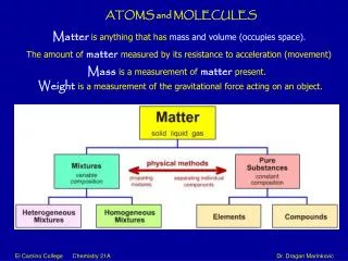
Mass Spectrometry Separates charged atoms or molecules according to their mass-to-charge ratio Frequently used for determination of molecular mass of proteins and nucleic acids and other macromolecules very accuratefastrequires very little material http://www.bmss.org.uk/what_is/whatisframeset.html
Basic steps in mass spectrometry: 1. Ionize the sample in an ion source Many methods are available to ionize molecules Molecules are brought into the gas phase either prior to or at the time of ionization The molecular ion can be fragmented, depending on the procedure use. This yields structural information about the molecule 2. Ions are repelled out of the ion source and accelerated towards an analyzer. Must chose whether to observe positive or negative ions. Neutral species are not observed. http://www.bmss.org.uk/what_is/whatisframeset.html
Types of mass spectrometry analyzers 1. Field analyzer: deflects ion depending on mass/charge ratio Field strength is scanned so ions hit the deflector at different times. 2. Time of flight analyzer: Measures the time it takes for the ionized molecule to reach the detector. This depends on the charge/mass ration http://www.bmss.org.uk/what_is/whatisframeset.html
Field Analyzer http://chipo.chem.uic.edu/web1/ocol/spec/MS1.htm
Time-of-Flight Analyzer s0 sa sd Detector v m/z E t = t0 + ta + td + tm http://ms.mc.vanderbilt.edu/tutorials/maldi/maldi-ie_files/frame.htm
There are many types of ionization methods 1. Electron ionization (EI) is the original method (still widely used) A high energy beam of electrons is generated at a heated filament Sample molecules in the gas phase pass through this beam.Electrons are stripped off and go to the positive “trap” Result is sample molecules that carry positive charge. Molecules often fragment. http://www.bmss.org.uk/what_is/whatisframeset.html
Most frequently used ionization methods for the analysis of macromolecules 1. MALDI: matrix assisted laser desorption ionization Dissolve the sample in a “matrix” that is allowed to crystallize on a stainless steel target. matrix material: 2,5 dihydroxybenzoic acid or sinapinic acid Use a pulsed laser (337 nm) to desorb the sample into the gas phase and ionize it. Ionization is usually by protonation or deprotonation Usually little fragmentation. Mass range is virtually without limit
+ MALDI hu Target Matrix Analyte can get resolution of 1 part in 50,000 by MALDI MS
4000 [M+H]+ [M+H]2+ 3000 Igg, 1.5 pmole on target Counts 2000 [M+H]3+ [2M+H]+ 1000 0 50000 100000 150000 200000 250000 300000 Mass (m/z) IgG in Sinapinic Acid
Mixture of 3 Proteins in Sinapinic Acid 8000 Bovine Insulin, MW 5733.5 Cytochrome C, MW 12360.1 Trypsinogen, MW 23981 Ins+ 6000 Counts Cyt.C+ 4000 2Cyt.C+ Cyt.C2+ Trg+ Ins2+ Trg2+ 2000 0 5000 10000 15000 20000 25000 Mass
V43I ACP mutant: 8522.4 example of MALDI MS Acyl Carrier Protein from E. coli V43I mutant plus unprocessed N-terminal Met: 8653.1 minor peaks are protein plus monovalent and divalent cations Unusual properties of the acyl carrier protein (ACP) was discovered to be due to a single mutation: V43I The change in molecular weight is evident in MALDI mass spec of the purified proteins wild type ACP 8508.4 part of sequence of E. coli Acyl Carrier Protein J. Biol Chem (1996) 271, 15905-
Peptide mapping Mass accuracy : 10 to 50 ppm 1607.06 748.09 1884.32 11500 12000 12500 13000 13500 Mass (m/z) 1815.13 1378.23 1502.02 1271.10 Peptide sequencing by MALDI-PSD 600 800 1000 1200 1400 1600 1800 2000 2200 Mass (m/z) Protein Identification by MALDI-MS: A strategy for proteomics 1-D blot Sample elution Sampling Mass of the protein Accuracy : 10-4 Separation on gel 2-D Digestion (specific protease) Database search Protein identification
Most frequently used ionization methods for the analysis of macromolecules 2. Electrospray ionization (ESI) Sample is dissolved in liquid, e.g., 1:1 water/methanol Squirted through a capillary held at very high potential at atmospheric pressure, generating a spray of charged droplets Nitrogen gas directs the droplets into chambers of increasing vacuum, causing loss of solvent and concentration of charge End up with sample molecules in multiple ionization states, but not fragmented
ESI of horse cytochrome c: multiple ionization states of a single molecule results in a complex pattern, but this can be mathematically analyzed and combined to reveal the mass of the sample molecule
Resolution available today is usually better than 1 part per 50,000 Tamoxifen MALDI ToF (time of flight)
1673.9 Average Mass 100 90 80 70 low resolution 1000 60 50 40 30 20 10 1,672 1,674 1,676 1,678 1,680 1,682 1672.9 Monoisotopic Mass [M+H]+ 100 90 [M+H+1]+ 80 70 [M+H+2]+ high resolution 10,000 60 50 40 [M+H+3]+ 30 20 [M+H+4]+ 10 0 1,672 1,674 1,676 1,678 1,680 1,682 Example of ESI MS: Neurotensin QLYENKPRRPYIL MW 1672.9
Tandem Mass Spectrometry (MSMS) Essentially two mass spectrometers in series. First spectrometer is used to select a designated ion, which is then directed to a collision chamber. Pressurized inert gas (Argon) is used to cause collision induced dissociation resulting in product ions (fragments) The product ions are then sent to the second analyzer MS-MS of 4-hydroxy Tamoxifen
Tandem MALDI ion trap mass spectrum of an “in-gel” trypsin digest of a 22 kDa protein from an SDS-PAGE gel MS of trypsin fragments Tandem MS of the m/z 2147.6 fragment Identification of the protein by matching peptide fragments with a bioinformatics library: gag protein HTLV-1 Anal. Chem.(1997) 69,3995-4001
Assignment of peptides of the gag HTLV-1 protein (22 KDa) to the trypsin fragment mass spectrum and confirmation by Tandem MALDI mass spectrum of the 229-240 fragment MALDI mass spectrum from an in-gel trypsin digest after SDS-PAGE Tandem MALDI of the m/z 1352.8 fragment, confirming its assignment as 229-240 of the gag protein. fragments yield sequence information