Download
1 / 99
1.03k likes | 1.35k Vues
Muscles of the Human Body. Science Olympiad Anatomy and Physiology 2009-2010. Muscles . Definition: a type of tissue composed of contractile cells (or fibers) which effect movement of an organ or part of the body Male and female body contains approximately 640 muscles.

E N D
Muscles of the Human Body Science Olympiad Anatomy and Physiology 2009-2010
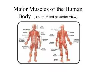
Muscles • Definition: a type of tissue composed of contractile cells (or fibers) which effect movement of an organ or part of the body • Male and female body contains approximately 640 muscles.
Skeletal Muscles • vary considerably in size, shape, and arrangement of fibers. • range from extremely tiny strands such as the stapedium muscle of the middle ear to large masses such as the muscles of the thigh. • may be made up of hundreds, or even thousands, of muscle fibers bundled together and wrapped in a connective tissue covering.
Characteristics of muscle • Excitability-responds to a stimuli (eg nerve impulse • Contractility-able to shorten in length • Extensibility-stretches when pulled • Elasticity-tends to return to original shape and length after contraction or extension
Functions of muscle • Motion: provide levers for muscles • The stimulation of individual muscle fibers maintains a state of muscle contraction known as tonus (muscle tone). • Important in maintaining the movement of blood and lymph through out the body. • When muscle is cut off from nerve supply, a condition that occurs when spinal nerves are severed, the muscles lose tonus and become flaccid and eventually atrophies (shrinks)
Heat production • Muscle metabolism produces heat as an end product. • Because muscles constitute about 40-45% of the body’s weight and are in a constant state of fiber activity, they are the primary source of body heat. • The rate of heat production rises with increased muscle activity. • Emaciated and elderly people, who have reduced muscle mass have difficulty staying warm.
Support: form the framwork that suports the body and cradles soft organs • Maintenance of posture • Skeletal muscles maintain posture, stabilize the joints and support the viscera. • Skeletal muscles have muscle tone (remain partly contracted), which helps maintain body posture. • Ongoing signals from the nervous system to the muscle cells help maintain tone and readiness for physical activity
Postural muscles of the head, neck and trunk are working even when you think you are relaxed. • The head, in particular is constantly being held at the atlanto-occipital joint up by the muscles of the neck. • When you start to get drowsy, these muscles will relax and your head nods forward.
Protection: provide a protective case for the brain, spinal chord, and vital organs • Mineral storage-reservoir for minerals especially calcium and phosphorus • Blood cell formation: hematopoiesis occurs within the marrow cavities of bones
Muscles • skeletal muscles will not contract unless stimulated by neurons • smooth & cardiac muscle will contract without nervous stimulation but their contraction can be influenced by the nervous system.
Types of muscle • skeletal: • attached to bones & moves skeleton • also called striated muscle (because of its appearance under the microscope, as shown in the photo to the left) • voluntary muscle Skeletal muscle (striated muscle)
Types of muscle • smooth • involuntary muscle • muscle of the viscera (e.g., in walls of blood vessels, intestine, & other 'hollow' structures and organs in the body) Smooth muscle
Types of muscle • cardiac • muscle of the heart • Involuntary • Myofibrils in the two interlocking muscle cells are firmly anchored to the membrane at the intercalated disc. • Because their myofibrils are essentially locked together, the two muscle cells can "pull together" with maximum efficiency. Cardiac muscle
Muscle attachments • Tendons: are dense connective tissue that attaches the muscle to bone. • When a muscle contracts, it shortens and puts tension on the tendon andthe bone. • The muscle tension causes movement of the bone at a synovial joint. • Belly: • the fleshy thick part of the muscle. Also called the gaster.
Origin • The less moveable attachment of the muscle • At the girdles and appendages the most proximal muscle attachment is the origin. • Insertion • The more moveable bony attachment of the muscle is called the insertion. • In muscles associated with girdles and appendages the more distal attachment is the insertion.
Muscle Groups based on their actions • Synergistic: • Muscle groups that contract together to accomplish a particular movement. • large movements of the body require several synergistic muscles to accomplish the task. • Muscles that are primarily responsible for a movement are called prime movers.
Muscle Groups based on their actions • Antagonistic: • muscles that have opposing actions and are located on opposite sides of a joint • are needed because the fibers in a contracted muscle are shortened and need to be elongated (stretched) before they can cause movement via contraction again.
Attached to bone by tendons composed of connective tissue The connective tissue surrounds the entire muscle and is called epimysium Skeletal muscle
Each muscle is surrounded by a connective tissue sheath called the epimysium. • Fascia, connective tissue outside the epimysium, surrounds and separates the muscles. • Portions of the epimysium project inward to divide the muscle into compartments. • Each compartment contains a bundle of muscle fibers.
Generally, an artery and at least one vein accompany each nerve that penetrates the epimysium of a skeletal muscle. • Branches of the nerve and blood vessels follow the connective tissue components of the muscle of a nerve cell and with one or more capillary
skeletal muscle • Skeletal muscles consist of many bundles called fascicles which are surrounded by connective tissue called Perimysium • Each fascicles is composed of numerous muscle fibers (or muscle cells) • The fascicles are surrounded by connective tissue called endomysium
Muscle cell • The cell plasma membrane of a muscle cell is called the sarcolemma, • maintains a membrane potential. • Forms a physical barrier against the external environment • Mediates signals between the exterior and the muscle cell
impulses travel along muscle cell membrane (sarcolemma) • the 'function' of impulses in muscle cells is to bring about contraction.
Sarcoplasm • Specialized cytoplasm of a muscle cell • Contains subcellular elements an the Golgi apparatus • Abundant with myofibrils • Has a modified endoplasmic reticulum known as the sarcoplasmic reticulum (SR) • Also has myoglobin and mitochondria
Transverse tubules (t-tubules) • Extends through the sarcolemma, through the muscle cell to the opposite side sarcolemma • Allows impulses to penetrate the cell and activate the SR (sarcoplasmic reticulum) • 2 tubules are in each sarcomere
Sarcoplasmic reticulum • A form of endoplasmic reticulum • Forms a network around the myofibrils • Stores and provides the Ca2+ that is required for muscle contraction
Myofibrils • Contractile units of the muscle cell • In an ordered arrangement of longitudinal myofilaments • Myofilaments are thick (myosin) or thin filaments (actin)
Muscle striations • The sarcomeres are what give skeletal and cardiac muscles their striated appearance. • A sarcomere is defined as the segment between two neighboring Z-lines (or Z-discs, or Z bodies). • Sarcomere’s line up end to end and work together for muscle contraction
Muscle striations • Surrounding the Z-line is the region of the I-band (for isotropic) • Area of thinner (lighter) filaments • A-band (for anisotropic) • Area of darker filaments
Within the A-band is a paler region called the H-band (from the German "Heller", bright).. • Finally, inside the H-band is a thin M-line (from the German "Mittel", middle of the sarcomere).
Myofibrils • Composed of 2 types of myofilaments • Thick-myosin • Thin-actin • Arranged in a very regular, précis pattern • Thick myofilaments are usually surrounded by 6 thin myofilaments • Anchoring structure is nebulin for the actin and titin for the myosin
http://highered.mcgraw-hill.com/sites/0072495855/student_view0/chapter10/animation__sarcomere_contraction.htmlhttp://highered.mcgraw-hill.com/sites/0072495855/student_view0/chapter10/animation__sarcomere_contraction.html
Myosin (thick filament) • Mysoin head • Has ATP binding sites in which ATP is housed • Has actin binding sites which fit molecules of ACTIN • Has a hinge at the point where it leaves the core of the thick filament, it swivels and actually causes muscle contraction
Actin (thin filament) • Composed of 3 types of protein: Actin, troponin, and tropomyosins wrapped around each other
http://highered.mcgraw-hill.com/sites/0072495855/student_view0/chapter10/animation__breakdown_of_atp_and_cross-bridge_movement_during_muscle_contraction.htmlhttp://highered.mcgraw-hill.com/sites/0072495855/student_view0/chapter10/animation__breakdown_of_atp_and_cross-bridge_movement_during_muscle_contraction.html
Nerve-muscle connection • The nervous system 'communicates' with muscle via neuromuscular junctions. • chemical transmitter is released from vesicles (each of which contains 5,000 - 10,000 molecules of acetylcholine) and diffuses across the neuromuscular cleft, • the transmitter molecules fill receptor sites in the membrane of the muscle & increase membrane permeability to sodium, • sodium then diffuses in & the membrane potential becomes less negative, • if the threshold potential is reached, an action potential occurs, an impulse travels along the muscle cell membrane, and the muscle contracts.
Steps in contraction of a muscle 1. Impulse is transferred from a neuron to the sarcolemma of a muscle cell 2. Impulse travels along the sarcolemma and travels down the T-tubules into the sarcoplasmic reticulum 3. impulse travels along the sarcoplasmic reticulum, opening the calcium gates in the membrane of the SR and allowing the calcium to diffuse out of the SR and among the myofilaments
Steps in contraction 4. Calcium fills the binding sites in the Troponin molecules which then alters the shape and position of the troponin and causes movement of the attached Troopomyosin molecule 5. Movement of the tropomyosin permits the myosin head to contact ACTIN 6. Contact with actin causes the myosin head to swivel
8. During the swivel, the MYOSIN HEAD is firmly attached to ACTIN. So, when the HEAD swivels it pulls the ACTIN (and, therefore, the entire thin myofilament) forward.. Many MYOSIN HEADS are swiveling simultaneously, or nearly so, and their collective efforts are enough to pull the entire thin myofilament).
8. At the end of the swivel, ATP fits into the binding site on the cross-bridge & this breaks the bond between the cross-bridge (myosin) and actin. The MYOSIN HEAD then swivels back. As it swivels back, the ATP breaks down to ADP & P and the cross-bridge again binds to an actin molecule. 9. As a result, the HEAD is once again bound firmly to ACTIN. However, because the HEAD was not attached to actin when it swiveled back, the HEAD will bind to a different ACTIN molecule (i.e., one further back on the thin myofilaments). Once the HEAD is attached to ACTIN, the cross-bridge again swivels,
Muscle fatigue • Under most circumstances, calcium is the "switch" that turns muscle "on and off" (contracting and relaxing). • When a muscle is used for an extended period, ATP supplies can diminish. • As ATP concentration in a muscle declines, the MYOSIN HEADS remain bound to actin and can no longer swivel. • This decline in ATP levels in a muscle causes MUSCLE FATIGUE. Even though calcium is still present (and a nervous impulse is being transmitted to the muscle), contraction (or at least a strong contraction) is not possible.
Types of muscle contractions • Isotonic- Contraction leads to shortening of the muscle. • involves movement against resistance and is a dynamic contraction. • Lifting free weights is primarily isotonic. and biceps curls are isotonic. • Isometric- is a static contraction in which the muscle remains the same length. • There is no shortening of the muscle • Usually performed against a resistance that can’t be moved