Download
1 / 166
1.68k likes | 2.11k Vues
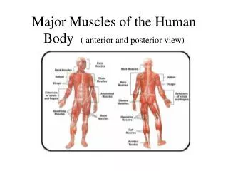
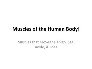
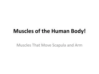
Muscles of the Human Body. Zack Zenko. Start. Navigation. Picture. Semitendinosus. Origin: ischial tuberosity Insertion: pes anserinus Innervation: sciatic nerve (tibial) Blood Supply: inferior gluteal artery, perforating arteries Function: flexes the knee, extends the hip .

E N D
Muscles of the Human Body Zack Zenko Start
Navigation Picture Semitendinosus • Origin: ischial tuberosity • Insertion: pes anserinus • Innervation: sciatic nerve (tibial) • Blood Supply: inferior gluteal artery, perforating arteries • Function: flexes the knee, extends the hip
Semitendinosus Navigation
Navigation Picture Soleus • Origin: upper half of the posterior surface of the tibia, upper third of the posterior fibula • Insertion: posterior surface of the calcaneus via the achilles tendon • Innervation: tibial nerve • Blood Supply: popliteal artery trunk, posterior tibial artery, peroneal artery • Function: plantar flexion
Navigation Soleus
Navigation Picture Flexor Digitorum Brevis • Origin: Calcaneus • Insertion:Phalanges of toe 2-5 • Innervation: medial plantar nerve • Blood Supply: medial and lateral plantar arteries • Function: flexes lateral four toe
Navigation Flexor Digitorum Brevis
Navigation Picture Abductor Hallucis • Origin: tuberosity of the calcaneus • Insertion: medial aspect of base of 1st phalanx of hallux • Innervation: medial plantar nerve • Blood Supply: medial and lateral plantar arteries • Function: abducts hallux
Navigation Abducts Hallucis
Navigation Picture Buccinator – The Trumpeter Muscle • Origin: Calcaneus • Insertion: fibers of the orbicularis oris • Innervation: buccal branch of the facial nerve • Blood Supply: buccal artery • Function: compresses the cheeks against the teeth and is used in blowing, assists the masseter in chewing
Navigation Picture Psoas Major • Origin: transverse processes of T12-L5, lateral aspects of intervertebral disks between them • Insertion:lesser trochanter of femur • Innervation: lumbar plexis • Blood Supply: lumbar branch of iliolumbar artery • Function: flexes and laterally rotates the thigh
Navigation Picture Iliacus • Origin: iliac fossa • Insertion: lesser trochanter of femur • Innervation: femoral nerve • Blood Supply: medial femoral circumflex artery, iliac branch of iliolumbar artery • Function: flexes and laterally rotates the thigh
Navigation Picture Obturator Externus • Origin: obturator foramen and obturatory membrane • Insertion: trochanteric fossa of femur • Innervation: posterior branch of obturator nerve • Blood Supply: obturator artery • Function: adducts thigh, laterally rotates thigh
Navigation Picture Obturator Internus • Origin: ischiopubic ramus and obturator membrane • Insertion: medial aspect of greater trochanter • Innervation: nerve to obturator internus; L5, S1 • Blood Supply: superior gluteal artery • Function: adducts thigh, laterally rotates thigh, stabilizes hip during walking
Navigation Picture Gemellus Inferior • Origin: ischial tuberosity • Insertion:obturator nternus tendon • Innervation: nerve to quadratus femorus; L4-S1 • Blood Supply: inferior gluteal artery • Function: laterally rotates thigh
Navigation Picture Gemellus Superior • Origin: spine of the ischium • Insertion: obturator internus tendon • Innervation: nerve to obturator internus; S1, S2, S3 • Blood Supply: inferior gluteal artery • Function: laterally rotates thigh
Navigation Picture Tensor Fasciae Latae • Origin: iliac crest • Insertion: iliotibial tract • Innervation: superior gluteal nerve; L4, L5 • Blood Supply: lateral circumflex femoral artery, superior gluteal artery • Function: flexes thigh, medially rotates the thigh, abducts thigh, stabilizes trunk
Navigation Picture Adductor Magnus • Origin: pubis, ischial tuberosity • Insertion: femur • Innervation: posterior branch of obturator nerve, sciatic nerve • Blood Supply: obturator artery • Function: adducts and extends hip
Navigation Picture Adductor Longus • Origin: pubic body just below the pubic crest • Insertion: middle third of linea aspera • Innervation: anterior branch of obturator nerve • Blood Supply: obturator artery • Function: adducts the thigh, flexes thigh
Navigation Picture Gluteus Maximus – The Butt Muscle • Origin: Gluteal surface of ilium, lumbar fascia, sacrum, sacrotuberus ligament • Insertion: gluteal tuberosity of femur, iliotibial tract • Innervation: inferior gluteal nerve; L5, S1, S2 • Blood Supply: superior and inferior gluteal arteries • Function: external rotation and extension of the hip, main antigravity muscle in sitting
Navigation Picture Gluteus Medius • Origin: Gluteal surface of ilium • Insertion: greater trochanter of femur • Innervation: superior gluteal nerve; L4, L5, S1 • Blood Supply: superior gluteal artery • Function: works with gluteus minimus, abducts hip and prevents adduction of the hip, medially rotates the thigh
Navigation Picture Gluteus Minimus • Origin: Gluteal surface of ilium • Insertion: greater trochanter of femur • Innervation: superior gluteal nerve; L4, L5, S1 • Blood Supply: superior gluteal artery • Function: works with gluteus medius, abducts hip and prevents adduction of the hip, medially rotates the thigh
Navigation Picture Pectineus • Origin: superior pubic ramus • Insertion: pectineal line of femur • Innervation: femoral nerve • Blood Supply: obturator artery • Function: flexes thigh, adducts thigh, medially rotates thigh
Navigation Picture Sartorius – The Tailor Muscle • Origin: inferior to the anterior superior iliac spine • Insertion: anteromedial surface of the upper tibia and pes anserinus • Innervation: femoral nerve • Blood Supply: femoral artery • Function: flexes, abducts, and laterally rotates the hip, flexes of the knee
Navigation Picture Gracilis • Origin: ischiopubic ramus • Insertion: tibia (pes anserinus) • Innervation: anterior branch of obturator nerve • Blood Supply: medial circumflex of femoral artery • Function: flexes, medially rotates, and adducts the hip, flexes the knee
Navigation Picture Semimembranosus • Origin: ischial tuberosity • Insertion: medial surface of tibia • Innervation: sciatic nerve; L5, S1, S2 • Blood Supply: profunda femoris, gluteal artery • Function: extends the hip, flexes the knee
Navigation Picture Biceps Femoris • Origin: ischial tuberosity, linea aspera, femur • Insertion: head of fibula • Innervation: long head: tibial nerve; short head: common peroneal nerve • Blood Supply: inferior gluteal artery, perforating arteries, popliteal artery • Function: flexes the knee, laterally rotates a flexed knee, and extends the hip
Navigation Picture Articularis Genu • Origin: femur • Insertion: suprapateller bursa • Innervation: femoral nerve • Blood Supply: femoral artery • Function: pulls the suprapateller bursa during knee extension
Navigation Picture Vastus Medialis • Origin: femur • Insertion: patella • Innervation: femoral nerve • Blood Supply: femoral artery • Function: extends the knee
Navigation Picture Vastus Lateralis • Origin: greater trochanter, intertrochanderic line, and linea aspera of femur • Insertion: patella via quadriceps tendon and tibial tuberosity via pateller ligament • Innervation: femoral nerve • Blood Supply: femoral artery • Function: extends and stabalizes the knee
Navigation Picture Rectus Femoris • Origin: anterior inferior iliac spine • Insertion: pateller tendon • Innervation: femoral nerve • Blood Supply: lateral femoral circumflex artery • Function: extends the knee, flexes the hip
Navigation Picture Vastus Intermedius • Origin: anterior lateral femur • Insertion: pateller ligament • Innervation: femoral nerve • Blood Supply: femoral artery • Function: extends knee
Navigation Picture Gastrocnemius • Origin: superior to articular surfaces of lateral condyle of femur and medial condyle of femur • Insertion: achilles tendon • Innervation: sciatic nerve; S1-S2 • Blood Supply: sural arteries • Function: plantar flexes foot, flexes knee
Navigation Picture Plantaris • Origin: lateral supracondylar ridge of femur • Insertion: achilles tendon • Innervation: tibial nerve • Blood Supply: sural arteries • Function: plantar flexes foot, flexes knee
Navigation Picture Soleus • Origin: fibula, medial border of tibia (soleal line) • Insertion: achilles tendon • Innervation: tibial nerve; S5 – S2 • Blood Supply: sural arteries • Function: plantar flexes foot
Navigation Picture Popliteus • Origin: middle facet of the lateral surface of the lateral femoral condyle • Insertion: posterior tibia under tibial condyles • Innervation: tibial nerve • Blood Supply: popliteal artery • Function: medially rotates and flexes the knee
Navigation Picture Peroneus Brevis • Origin: fibula • Insertion: fifth metatarsal • Innervation: superficial peroneal nerve • Blood Supply: peroneal artery • Function: plantar flexes foot, everts of the foot
Navigation Picture Peroneus Longus • Origin: fibula • Insertion: first metatarsal, medial cuneiform • Innervation: superficial peroneal nerve • Blood Supply: peroneal artery • Function: plantar flexes foot, everts of the foot
Navigation Picture Tibialis Posterior • Origin: fibula, tibia • Insertion: navicular, medial cuneiform • Innervation: tibial nerve • Blood Supply: posterior tibial artery • Function: plantar flexes foot, inverts of the foot
Navigation Picture Tibialis Anterior • Origin: body of tibia • Insertion: first metatarsal, medial cuneiform • Innervation: deep peroneal nerve • Blood Supply: anterior tibial artery • Function: dorsiflexes foot, inverts the foot
Navigation Picture Masseter – The Chewing Muscle • Origin: zygomatic arch and maxilla • Insertion: coronoid process and ramus of mandible • Innervation: mandibular nerve • Blood Supply: masseteric artery • Function: opens and closes the mouth
Navigation Picture Zygomaticus Major • Origin: zygomatic bone • Insertion: modiolus of mouth • Innervation: zygomatic branch of facial nerve • Blood Supply: facial artery • Function: draws angle of mouth upward and laterally
Navigation Picture Zygomaticus Minor • Origin: zygomatic bone • Insertion: skin of the upper lip • Innervation: buccal branch of facial nerve • Blood Supply: facial artery • Function: elevates upper lip
Navigation Picture Orbicularis Oris – The Kissing Muscle • Origin: maxilla and mandible • Insertion: skin around the lips • Innervation: buccal branch of facial nerve • Blood Supply: facial artery • Function: puckers the lips
Picture Navigation Orbicularis Oculi – The Winking Muscle • Origin: frontal bone, medial palpebral ligament, lacrimal bone • Insertion: lateral palpebral raphe • Innervation: zygomatic branch of facial nerve • Blood Supply: ophthalmic artery, zygomatico-orbital artery • Function: closes the eyelids
Navigation Picture Frontalis • Origin: galea aponeurotica • Insertion: mastoid process • Innervation: facial nerve • Blood Supply: ophthalmic artery • Function: wrinkles eyebrow
Navigation Picture Levator Scapulae • Origin: posterior tubercles of transverse processes of C1 – C4 • Insertion: superior part of medial border of scapula • Innervation: cervical nerves; C3, C4 and dorsal scapular nerve; C5 • Blood Supply: dorsal scapular artery • Function: elevates scapula and tilts its glenoid cavity inferiorly by rotating scapula
Navigation Picture Scalene Muscles • Origin: cervical vertebrae; C2 – C7 • Insertion: first and second ribs • Innervation: cervical nerves; C3 – C6 • Blood Supply: ascending cervical artery • Function: elevates ribs 1 and 2
Navigation Picture Sternocleidoidmastoid • Origin: manubrium sterni, medial portion of clavical • Insertion: mastoid process of temporal bone, superior nuchal line • Innervation: accessory nerve, cervical plexus • Blood Supply: occipital artery, superior thyroid artery • Function: on its own – tilts head towards its own side and turns face to opposite side; in unison – flexes neck, raises sterum, assists in forced inspiration
Navigation Picture Extensor Digitorum • Origin: lateral epicondyle • Insertion: extensor expansion of the middle and distal phalanges of fingers 2 – 5 • Innervation: posterior interosseus nerve • Blood Supply: posterior interosseus artery • Function: extends the hand, wrist, and fingers