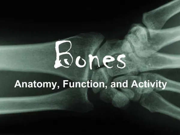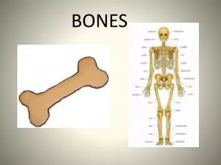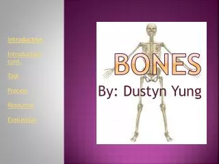
BONES
BONES. By Dr.PARDEEP KUMAR. Bones. Bone is a highly vascularised, mineralised & rigid type of connective tissue. Composition of Bone. Bone cells Osteoproginator cells, Ostoeoblast , Osteoclast , Osteocytes Intercellular matrix Organic Collagen Fibers Glycosaminoglycans

BONES
E N D
Presentation Transcript
BONES By Dr.PARDEEP KUMAR
Bones • Bone is a highly vascularised, mineralised & rigid type of connective tissue.
Composition of Bone • Bone cells • Osteoproginator cells, Ostoeoblast, Osteoclast, Osteocytes • Intercellular matrix • Organic • Collagen Fibers • Glycosaminoglycans • Glycoprotein • Proteo-glycans • Inorganic • Mostly Calcium phosphate • Calcium carbonate • Magnesium hydroxide, fluoride & sulphate (small amount
Functions of Bone • Bones perform several important functions: • Support • Protection • Movement • Mineral storage • Blood cell formation
Function of bones • Support • Bones provide a hard framework that gives shape and support to the body . • Bones provide support for internal organs.
Function of Bones • Protection • Fused bones provide a brain case that protects this vital tissue • Spinal cord is surrounded by vertebrae • Rib cage protects vital organs
Function of Bones • Movement • Skeletal muscle attached to bones use the bones as levers to move the body • Arrangement of bones and joints determine the movements possible
Function of Bones • Mineral storage • Bone serves as a mineral reservoir • Phosphate and calcium ions can be released into the blood steam for distribution • Deposition and removal are ongoing
Function of Bones • Blood cell formation • Hematopoiesis occurs within the marrow cavities of the long bones. • The majority of hematopoiesis occurs in bones.
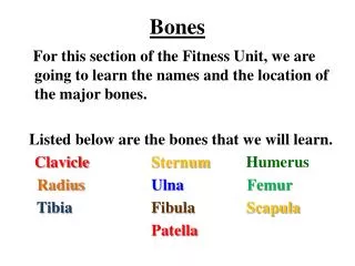
Classification of Bones (According to structure) • Compact bone:- shaft of long bone • Spongy bone:- ends of long bone covered by thin layer of compact bone ( According to size & shape) • Long bone – femur, tibia, fibula. • Short bone – Carpal & tarsal bones • Irregular bone – Vertebrae, Hip bones • Flat Bone – Sternum, , Parietal, Frontal • Sesamoid bone – Patella, pisiform • Pneumatic Bone – Maxilla
Classification According to size & shape • Long bones • Typically longer than wide • Have a shaft with heads at both ends • Contain mostly compact bone • Examples: Femur, humerus • Short bones • Generally cube-shape • Contain mostly spongy bone • Examples: Carpals, tarsals
Classification According to size & shape • Irregular bones • Irregular shape • Do not fit into other bone classification categories • Example: Vertebrae and hip • Flat bones • Thin and flattened • Usually curved • Thin layers of compact bone around a layer of spongy bone • Examples: Skull, ribs, sternum
Classification According to size & shape • Pneumatic Bone • Some bones of base of skull contain air-filled cavities called air sinuses or air cells. • These bones are called pneumatic bones. • These cavities make the skull lighter and gives resonance to the voice. Examples: Maxilla Ethmoid Mastoid. • Sesamoid Bones • These are bony nodules located in the region where the tendon crosses a joint. • Functions: • These bones function as a fulcrum. • They serve to protect the tendons by limiting trauma from friction or pressure. Examples: • Patella or knee cap
Outer layer of bone. • It is very hard and dense. • It contains cylinders of calcified bone called osteons. • Osteon provides great strength needed to resist the stress to which long bones are subjected. • Located in the ends of long bones. • Many spaces that are filled with red bone marrow which produces bone cells. • It provides great strength with least weight.
Microscopically the structure of spongy & compact bone is the same • The only difference is that the space in the spongy bone is large while in the compact type the bone tissue is dense & contains narrow channels
Diaphysis • Shaft • Composed of compact bone • Epiphysis • Ends of the bone • Composed mostly of spongy bone Gross Anatomy of a Long Bone Figure 5.2a
Periosteum • Outside covering of the diaphysis • Fibrous connective tissue membrane • Endosteum • It is highly vascular membrane which lines the medullary cavity in the inner surface of compact bone • Sharpey’sfibers • Secure periosteum to underlying bone • Arteries • Supply bone cells with nutrients Structures of a Long Bone
Articular cartilage • Covers the external surface of the epiphyses • Made of hyaline cartilage • Decreases friction at joint surfaces Structures of a Long Bone Figure 5.2a
Medullary cavity • Cavity of the shaft • Contains yellow marrow (mostly fat) in adults • Contains red marrow (for blood cell formation) in infants Structures of a Long Bone Figure 5.2a
Osteon (Haversian System) • A unit of bone • Central (Haversian) canal • Opening in the center of an osteon • Carries blood vessels and nerves • Perforating (Volkman’s) canal • Canal perpendicular to the central canal • Carries blood vessels and nerves Microscopic Anatomy of Bone
Microscopic Anatomy of Bone Figure 5.3
Lacunae • Cavities containing bone cells (osteocytes) • Arranged in concentric rings • Lamellae • Rings around the central canal • Sites of lacunae Microscopic Anatomy of Bone Figure 5.3
Microscopic Anatomy of Bone • Canaliculi • Tiny canals • Radiate from the central canal to lacunae • Form a transport system Figure 5.3
Blood supply of bones • Bone has a rich supply of blood. The distribution varies according to the type of bone Long Bone Long bone is supplied by 4 sets of blood vessels • Nutrient artery • Juxta-Epiphyseal or Metaphyseal vessels • Epiphyseal vessels • Periosteal Vessels
Nutrient artery • Enters the shaft through the nutrient foramen obliquely & reaches the medullary cavity where it is divided into ascending & descending branches Area of supply • Medullary cavity, inner 2/3rd of compact bone of Diaphysis & metaphysis
Juxta-Epiphyseal or Metaphyseal vessels • These are derived from the anastomosis around the joint Area of Supply • They supply the Epiphyseal end Epiphyseal vessels • Derived from the anastomosis around the joint Area of supply • The spongy part & red marrow of bones Periosteal Vessels • Derived from the vessels of periosteum Area of supply • Outer 1/3rd of compact bone
Blood Supply of short Bones • Recieve numerus fine blood vessels from the periosteum • These vessels enter through non-articular surface • Supply both compact and spongy bone, & bone marrow.
Blood Supply of Flat Bones • Periosteal plexus. Blood Supply of Irregular Bones • Large nutrient artery • Periosteal plexus.
Nerves Of Bone • Nerves are most numerus at articular areas of all types of bone. • Nerves are distributed throughout the periosteum. • Periosteum is the most sensitive part. • Compact bone is less sensitive to spongy bone.
Ossification • The process of gradual bone formation by depositing minerals from membranous or cartilaginous model is called ossification Types of Ossification Intra membranous Ossification: • Involves the replacement of sheet like connective tissue membranes with bony tissue • In this process the future bones 1st formed as connective tissue membranes. Osteoblasts migrate to the membranes and deposit bony matrix around them. Examples of bones formed in this manner are certain flat bones of the skull and some irregular bones.
Intra cartilaginous: Involves the replacement of hyaline cartilage with bony tissue • In this process the future bones 1st formed as cartilage models. • They become infiltrated with blood vessels and Osteoblast. • The Osteoblast forms a collar of compact bone around the Diaphysis. • At the same time Osteoclast breakdown the central bone tissue to form the medullary cavity • Later usually after birth secondary centre of ossification develop in the epiphysis. • Ossification in the epiphysis is similar to that in the Diaphysis except that the spongy bone is retained instead of being broken down into medullary cavity.
Factors affecting growth of bone • Adequate blood supply • Availability of adequate amount of minerals esp. Ca & PO4 • Vitamin D – Essential for absorption, utilization & maintenance of Ca & PO4 at normal levels • Vitamin C – Essential for the normal matrix formation • Mechanical factors – Tensile force helps in bone formation . • Hormones • Growth Hormone & thyroid hormone [Thyroxine & triiodothyronine] – are especially important during infancy & childhood for development of skeleton. • Testosterone & Oestrogen – Influences the physical changes that occur at Puberty. • Calcitonin & Parathormone – are involved in Homeostasis of blood & bone calcium levels required for bone development.
Sinuses • Sinuses are small air filled cavities in the bone & are found in Frontal, Sphenoid, Ethmoid & maxillary.
Fontanel– • An area of unossified area lying between the cranial bones of the a foetus • E.g. Anterior Fontanel, Posterior Fontanel
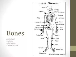
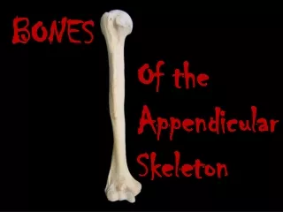
Skeleton The skeleton is described into 2 parts • Axial skeleton • Appendicular skeleton
Axial skeleton It consists of • Skull • Vertebral Column • Sternum • Ribs
Appendicular skeleton It consists of • Bones of Upper Limb & Shoulder • Clavicle • Scapula • Humerus • Ulna • Radius • Carpals [Scaphoid, Lunate, Triquetral, Pisiform, ] • [Trapezium, Trapezoid, Capitate, Hamate,] • Metacarpals • Phalanges
Bones of Lower Limb & Pelvis • Hip Bone • Femur • Tibia • Fibula • Patella • Tarsals • Metatarsals • Phalanges
Vertebral Column It consists of • 7 Cervical [Atlas, Axilla] • 12 Thoracic • 5 Lumbar • 1 Sacrum [5 fused bone] • 1 Coccyx [4 fused bone] • The bodies of the bones are separated from each other by intervertebral discs consisting of Cartilage.
Thoracic Cage Is formed by • 12 thoracic Vertebrae • 12 pairs if Ribs • 1 Sternum