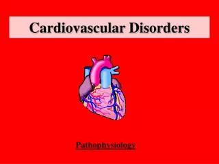
Physical therapy evaluation for cardiovascular disorders
230 likes | 450 Vues
Physical therapy evaluation for cardiovascular disorders. Ahmad Osailan. Cornerstone of cardiac assessment . Review of history Physical examination Diagnostic tests Functional capacity assessment . Review history . Risk factors: - Age, Smoking

Physical therapy evaluation for cardiovascular disorders
E N D
Presentation Transcript
Physical therapy evaluation for cardiovascular disorders Ahmad Osailan
Cornerstone of cardiac assessment • Review of history • Physical examination • Diagnostic tests • Functional capacity assessment
Review history • Risk factors: - Age, Smoking - High blood cholesterol (Dyslipidemia, Hyperglycemia) - High Blood pressure - Diabetes Millitus - obesity and overweight - Life style Activity - Past Medical History: History of MI or angina, dysarrythmias, artificial pacemaker.
Physical examination • 1) General examination: • - Cyanosis • - Pale face • - Dyspnea during conversation or with minimal activity • - Nutritional status: Malnutrition, obesity, overweight • - Skeletal deformities • - Tremor
Physical Examination • 2) Vitals: • - Heart rate • - Blood Pressure • - Pulse strength ( carotid A, radial A,) • - Breathing Rate ( tachypnea, Bradypnea) • - Breathing Pattern (shallow, Normal) • 3) Palpation • - Edema ( generalized or local) ( pitting or non pitting)
Physical Examination • 4) Auscultation: • Normal sound of the heart: S1 and S2 • ‘ Lubb = S1: sound of closure of AV valves • ‘ Dubb = S2 sound of closure of semilunarValves • http://www.youtube.com/watch?v=39n4XWv7flQ • - Presence of mumurs( abnormal sound of closure of valves) • - Breathing Sound ( different lubes) wheezing or cripitions may indicate pulmonary Edema
Diagnostic Tests • Types of Diagnostic Tests for cardiac disease: • Electrocardiogram (ECG) • Echocardiogram • Chest X- ray • Angiography
Diagnostic tests • Echocardiogram: • a sonogram of the heart. uses standard two-dimensional, three-dimensional, and Doppler ultrasound to create images of the heart. • What we care about in Echo: • Early filling and atrial filling E/F ratio • Report of the Echo • Ejection fraction percentage.
Diagnostic Test • Chest X-ray: is a projection radiograph of the chest used to diagnose conditions affecting the chest, its contents, and nearby structures. • What do we care about chest X-ray: • Lubes of the lung • Any presence of pleural effusion • Any hypertrophy or change in size of heart
Diagnostic Test • Angiography: is a medical imaging technique used to visualize the inside, or lumen, of blood vessels and organs of the body, with particular interest in the arteries, veins and the heartchambers • http://www.youtube.com/watch?v=kY5gKdFWT3k
Diagnostic Tests • Electrocardiograph: A device used to detect electrical activity of the heart over a period of time. • The ECG allows observation of the heart electrical activity by visualizing waveform beat origin from SA node down to purkinje fibres. • Types of ECG: • 12 leads ECG • 5 Leads ECG • 3 Leads ECG
Diagnostic Tests • Electrocardiograph
Diagnostic Tests • Components of ECG: • P wave: represent atrial excitation • P R interval: represent AV node delay • QRS complex: marks ventricular Excitation • ST segment: shift in depression represent ischemic heart disease • T wave: marks repolarization. • http://www.youtube.com/watch?v=iGlxCtU4Ejw
Examples of ECG • Try to find out what is missing • Atrialfibrilation • http://www.youtube.com/watch?v=K_uccmtCqZI
Examples of ECG • Try to find out what is missing • Premature ventricular contraction PVC • http://www.youtube.com/watch?v=s7cJyoaM-Yg
Examples of ECG Premature atrial contraction PAC http://www.youtube.com/watch?v=HrhLtDdxAWc
Examples of ECG • Ventricular ectopic tachycardia • http://www.youtube.com/watch?v=i-_clh3oFwE
Functional capacity assessment • It is a fundamental requirement for ADL • Definition: Is the ability to perform aerobic, oxygen work exercise . • Such activities require high efforts from heart, lung. • Thus, assessment utilizes mostly oxygen consumption VO2 • Functional capacity is identified by METs
METs • Metabolic Equivalents: • is defined as the ratio of a person's working metabolic rate relative to the resting metabolic rate. • One MET represents the oxygen consumption of a resting adult (3.5 ml/kg/min) • Functional capacity is defined as :poor (<4 METS),moderate (4–7METS), good (>7–10METS)
Functional capacity assessment Through : Cardiopulmonary exercise testing (stress test) Incremental Shuttle Walking Test. Chester step test 6 MWT