Physical Therapy for Cardiovascular disorders
640 likes | 872 Vues
Physical Therapy for Cardiovascular disorders. RHPT 482 Credit hours: 2T+1C Course Instructor: Ahmad Osailan. Course Description . The course is designed to teach and perform clinical practice for the management of CVD. Anatomy and physiology of the cardiovascular system. Objectives :

Physical Therapy for Cardiovascular disorders
E N D
Presentation Transcript
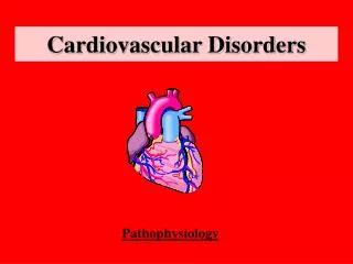
Physical Therapy for Cardiovascular disorders RHPT 482 Credit hours: 2T+1C Course Instructor: Ahmad Osailan
Course Description • The course is designed to teach and perform clinical practice for the management of CVD.
Anatomy and physiology of the cardiovascular system • Objectives : • Be familiar with the anatomy of the heart and vascular system • Size of the heart • Location of the heart • Layers of the heart • Chambers of the heart • Vascular system and its layers • Brief physiology of cardiovascular system
Cardiovascular system • Consist of : • Heart • Blood vessels • Lymphatic
Overview of the heart • Heart Definition: “a pump that moves the entire body volume to and from the lungs and tissues.” • In Humans, Heart is 250 g in Male to 350 in Female. (Size of a fist) • It produces ~ 5 Litres of Blood every minute • Myogenic: ability to generate its own contraction
Location of the heart • posterior to sternum • medial to lungs • anterior to vertebral column • base lies beneath 2nd rib • apex at 5th intercostal space • lies upon diaphragm
posterior to sternum • medial to lungs • anterior to vertebral column • base lies beneath 2nd rib • apex at 5th intercostal space • lies upon diaphragm
Heart Structure • Consist of : • Pericardium • Walls of the heart • Four champers • Four valves
Covering of the Heart • Pericardium: • Def: Is a fibrous sac surrounds the heart and roots of great vessels. • Divided to : • Serous pericardium: smooth inner • Fibrous pericardium: Tough fibrous tissue outer
Pericardium • Serous pericardium • Parietal layer: lines the inside of fibrous pericardium • Visceral layer: adheres to the surface of the heart. • Fibrous pericardium: Protects the heart and serous membrane
Heart Wall layers • Epicardium: outer layer • Myocardium: Middle layer • Endocardium: Inner layer
Endocardium • Inner lining • Smooth (Endothelial) surface that permits blood to move easily through the heart • Continuous with lining of blood vessels
Myocardium • Middle layer made of cardiac muscle (Myocardium) • Forms the bulk of the heart wall • Contains the septum- a thick muscular wall that completely separates the blood in the right side of the heart from the blood in the left side.
Epicardium • Protective, outer layer of the heart wall • same as the visceral pericardium • The coronary blood vessels that nourish • the heart wall are located here
Champers of the Heart • Heart has Four Champers • 2 atria (atrium) 2 ventricles • 2 atria separated by interatrial septum • 2 ventricles separated by interventricular septum.
Champers Right Atrium • Thinner wall than ventricles • Receives deoxygenated blood from vena cava • Passes blood through tricuspid valve into right • ventricle
Right Ventricle • Thicker wall than atria • Comprises most of anterior surface of heart • Circulates deoxygenated blood to lungs through the pulmonic valve into pulmonary trunk
Left atrium • Receives freshly oxygenated blood from pulmonary vein • Passes blood to left ventricle through mitral valve
Left ventricle • Receives blood from left atrium • Thickest myocardial wall • Forms apex of heart • Sends blood to systemic circulation via aorta
Valves of the heart • Function- prevent blood from flowing backwards • Responds to changes in pressure • Two types of valves in heart • Atrioventricular valves (AV) • Semi-lunar valves
Semilunar valves • Located at exit of ventricles, originiate from endothelial lining of veins • Heart contains two semilunar valves • Pulmonic • Aortic (Frequently damaged by Htn)
Atrioventricular Valves • Valve cusps are connected to papillary muscles • Chordaetendineae tiny collagen cords • that anchor cusps of valve to papillary muscles
Atrioventricular Valves • Left AV valve (Mitral, bicuspid) • Contains 2 cusps • Subject to abuse • Right AV valve (Tricuspid) • Contains 3 cusps • Not subjected to great abuses
Blood circulation • There are 2 circulatory pathways: • Pulmonary circuit • Systemic circuit
Blood circulation • Pulmonary circuit: Deoxygented Blood received by Right Atrium from Superior and inferior vena cava-> pass through Tricuspid V-> Right Ventricle -> through Pulmonary V -> pulmonary trunk -> pulmonary arteries -> R + L Lung.
Blood Circulation • Systemic Circuit: Oxygenated blood returns from lungs to heart through -> pulmonary veins -> Left Atrium -> pass through Mitral V -> Left ventricle -> pass through Aortic V -> aortic arch -> whole body
Blood Vessels • Function: • Distribution of blood • Exchange of materials with tissues • Return of blood to heart • Structure: • Most have 3 layers surrounding a hollow lumen
Blood Vessels • Arteries Veins • Arteriole Venule • Capillary network
General structure of Blood Vessels • Arteries and Veins Have 3 Tunics: • Tunica Externa ( Adventetia ) • Tunica Media • Tunica Intema • Capillaries composed of endothelium ( tunica intema)
Blood vessels • Tunica Intema: • Inner smooth layer • Simple squamous epithelium • Continous with endocardium • Present in all vessels
Blood Vessels • Tunica Media: • Layer of smooth Muscles • Contain Elastin • Supplied by Sympathetic division of ANS • Area of vasoconstriction and Vasodilatation
Blood Vessels • Tunica Externa • Layer of Connective tissue • Elastic and has collagen fibres
Arteries • Characteristics: • Thick walled • Lots of Elastin in all tunics • Stretchable wall to recoil and propel blood • Withstand and regulate BP Fluctuations
Veins • Characteristics: • Thin walled • Lumen is larger than arteries • Less stretchable than arteries
Capillaries • Characteristics : • Smallest vessel • Large enough for 1red cell • One tunica only (tunica intema) • Very thin, Why?
Coronary arteries • 2 Main Coronary Arteries •Right CA- branches into some marginal arteries; supplies RV and posterior of heart •Left CA- branches into • AIA (LAD) and • circumflex; supplies LV
Arterial supply to heart • Originates from the base of aortic artery • Only 5% of ejected blood is received through to innervate the heart (Myocardium) • Many branches directed to the Left ventricle, Why? • Coronary arteries traverse the heart forming a vast network of capillaries
Venous drainage • Transport deoxygenated blood to coronary sinus • Coronary Sinus drains into RA
Discussion • Which chamber among ventricles is thicker? • Which one is thicker among blood vessels? • How many layers surrounding the heart? • How many valves and what is the type of each one? • How many Circuit in cardiovascular system?
Mainly the heart is a pumping machine. • To pump it require force to generate contraction. • Since the heart is mainly composed of cardiac muscles (myocyte), • it has similar functions and structure like skeletal muscles with some variations
Myocyte • Characteristics: • Striated cells • Short cells • Mononucleus (one nucleus only) • Very large mitochondria (Many ATP) • Has intercalated disks