Cardiovascular Disorders
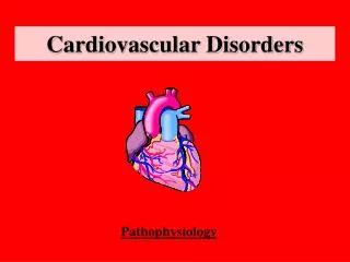
Cardiovascular Disorders. Chapter 5. Anatomy & Physiology. Four chamber Two atria Two ventricles. Chambers. Chambers Right and left atria Right and left ventricle. Valves . Conduction System. SA node is pacemaker Normal pacemaker is 60-100 Bradycardia <60 Tachycardia >100.

Cardiovascular Disorders
E N D
Presentation Transcript
Cardiovascular Disorders Chapter 5
Anatomy & Physiology • Four chamber • Two atria • Two ventricles
Chambers • Chambers • Right and left atria • Right and left ventricle
Conduction System • SA node is pacemaker • Normal pacemaker is 60-100 • Bradycardia <60 • Tachycardia >100
Cardiac Cycle Closure of tricuspid valves Closure of AV valves
General Evaluation(taken sitting, supine and lateral recumbent) • Aortic Valve – 2nd R intercostal space, right sternal border • Pulmonic Valve – 2nd L intercostal space, left sternal border • 2nd Pulmonic Valve (Erb’s Point) – 3rd intercostal space at left sternal border • Tricuspic Valve – 4th intercostal space along lower left sternal border • Mitral Valve: 5th intercostal space at apex of heart
Assessment Areas Aortic Pulmonic Erb’s Tricuspid Mitral
Auscultate the Precordium with Diaphragm & Bell • Note the rate and rhythm • Identify S1 and S2 • Extra heart sounds • S3 • S4 • Pericardial friction rubs
Lub Dup Lub Dup Lub Dup S1 & S2 • Identify S1 (heard best at apex) • S1 signals the beginning of systole • Identify S2 (heard best at base) • S2 signals the beginning of diastole • Pause after S2
S3 • Heard after S2 • Lower pitch than S2 • Heard better with the bell • Heard best at the apex of heart lub duppa lub duppa
S4 • Heard immediately before S1 • Lower pitch than S1 • Heard better with bell • Heard best at apex of heart daLub dup daLub dup
Murmurs • Restricted forward flow thru stenotic valve • Backward Flow thru regurgitant valve • Abnormal open in chambers
Six Characteristic of Murmurs • Timing • Loudness • Pitch • Pattern • Quality • Location
Cardiovascular Adaptations to Exercise • What happens to: • Cardiac Output (CO = HR X SV) • Stroke volume • Heart rate (both resting and max) • Venous return • Contractibility • Max VO2 • Heart size
Cardiovascular Adaptations to Exercise • What happens during maximal dynamic exercise? • CO increases 4-6X • Heart rates can triple • Stroke volume triples • # of capillaries • Blood flow to extremities
Sudden Cardiac Death • An unexpected and nontraumatic event that occurs instanteously or within minutes of an abrupt change in a person's previous clinical state.
Epidemiology • Over 5 million participants in sports with fewer than 20 individuals who die from sudden death syndrome (SDS) • Most are cardiac related and individuals are usually physically fit
Epidemiology • For SDS • 62% in HS • 22% in College • 9% middle school • 7% Professional • Young, male caucasions are the highest risk group in HS athletes • More prominent in weekend warriors
Epidemiology • Sports with “projectile” objects have a higher pre-disposition: • Baseball • Softball • Hockey • Lacrosse • Soccer • Football • Karate
Epidemiology • < 30 YO Hypertrophic Cadiomyopathy • Time of Death 3:00-9:00 PM • August-January most common months • > 30 YO Cardiovascular Disease • Usually in AM hours
Causes of SDS • HCM ( over 50% of all cases) • Coronary artery anomalies • Increased cardiac mass • Blunt trauma to chest cavity (commotio cordis)
Hypertrophic Cardiomyopathy (HCM) • Hypertrophy of L ventricle • No other CHD issues • Obstruction to ventricular outflow 20-30% of all cases • Occurs with Valsalva event • After exercise • Sudden change in posture • Diastolic dysfunction (80%)
HCM • Generally no previous symptoms prior to death • S/S • Syncope • Dyspnea • Fatigue • Angina (Chest pain) • Systolic ejection murmur
HCM DX • Left ventricle thickness of > 20 mm • Left ventricle outflow obstruction > 50 mm Hg • Family history of SDS (>40 YO) • Atria or ventricular arrhythmia
Diagnostics • Electrocardiogram – 90% accurate but does not detect ventricular outflow obstruction • Echocardiogram – Best Test • Secondary to arrhythmias • Most individuals are asymtomatic prior to death • IF HCM is detected, mortality rates are quite low (2 – 6%)
Coronary Artery Abnormalities Aberrant Coronary Arteries - 2 Types • Aberrant origin of the left coronary artery from the right sinus • Origin of the right coronary from the left sinus, with the anomalous artery coursing between the aorta and the pulmonary artery
Marfan’s Syndrome • ~40,000 individuals have Marfan’s in the US ( 1 in 10,000) • Genetic disorder that decreases the integrity of the connective tissues of the body • 30% of cases have no family history • 50% probability of passing to children from parents with Marfan’s syndrome • No ethnic tendency
Problems and Concerns • Mitral value prolapsed • Aorta is weak and dilated for inefficient blow flow • Aorta dissection
Physical Traits • Pes planus • Tall and thin body structure • Disproporante or unusually large appendages • Arm span greater than standing height • Hyperextensible joints or an elongated thumb • Scoliosis
Physical Traits • Convex/concave sternum (pigeon chest) • Genu recurvatum • Myopia • Abnormally enlarged heart • Increased incidence of hernia • Optic lens displacement
Clinical Tests • Thumb Sign • Wrist Test
Myocarditis • Caused by enteroviral infections (coxsackievirus B) • S/S • Typical viral responses (fever, nausea, etc) • Previous episodes of congestive heart failure • Fatigue, dyspnea, syncope, palpitations • Diagnostics - ECG
Congenital Aortic Stenosis • Bicuspid valve malformation • Impaired L ventrical outflow • Hypertrophy of interventricular septum and left ventricular (LV) free wall • Rheumatic heart disease • Atherosclerosis • S/S • Myocardial ischemia, LV dysfunction • Hypotension and exertional syncope
Mitral Valve Prolapse • Seen in 6 – 8% of pop • Possible cause of exercise induced SDS • S/S • Palpitations, shortness of breath • RTP • Asymptomatic athletes are able to participate – watch for warning signs (page 113)
Systemic Hypertension • BP > 140/90 • No distinguishable cause • S/S • Take BP on 3 separate occasions to confirm • Tx • Reduce alcohol consumption, meds, reduce weight
Deep Vein Thrombosis • Blood clot usually in vein of lower leg (however can occur anywhere in body) • Causes – trauma from surgery • 50% incidence rate after knee/hip replacement • Prolonged sitting (truckers, bus drivers) • Women who smoke, use oral contraceptives
Deep Vein Thrombosis cont. • S/S • Limb pain and edema • Homan’s Sign • Fever • Tx • Anticoagulants (Heparin, Coumadin)
Anemia • Low RBC count, decreased hemoglobin concentration • 3 types – iron deficiency, exertional hemolysis, dilutional pseudoanemia • S/S • Fatigue, tachycardia • Diagnosis – CBC • < 14 g/dl for men • < 12 g/dl for women
Sickle Cell Anemia • Genetic defect in hemoglobin common in African Americans • Anemia or Trait? Those with trait can participate in athletics, those with anemia cannot • Inability for O2 to bind with RBC • Red Flags (p 123)