Download
1 / 63
640 likes | 914 Vues
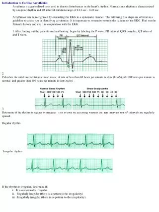
Management of Arrythmias According to AHA Guidelines. Dr Omar SCM Pharmacology. Common Arrythmias:. Asystole PEA Bradycardias Sinus 1 st Degree Heart Block 2nd Degree Heart Block Mobitz Type 1 Mobitz Type 2 3rd Degree Heart Block Tachycardias Narrow Complex Broad Complex.

E N D
Management of Arrythmias According to AHA Guidelines Dr Omar SCM Pharmacology
Common Arrythmias: • Asystole • PEA • Bradycardias • Sinus • 1st Degree Heart Block • 2nd Degree Heart Block • Mobitz Type 1 • Mobitz Type 2 • 3rd Degree Heart Block • Tachycardias • Narrow Complex • Broad Complex
Causes: H’s and T’s • Hypoxia (CNS events) • Hypokalemia/hyperkalemia (and other electrolytes) • Hypothermia/hyperthermia • Hypoglycemia/hyperglycemia • Hypovolemia (tank/anaphylaxis, gravid)
Causes: H’s and T’s (cont’d) • Trauma • Tamponade • Thrombosis (pulmonary) • Thrombosis (coronary) • Tablets (ODs, drugs, etc) • Tension (pneumothorax, asthma)
Asystole Algorithm (cont’d) Transcutaneous pacing: If considered, perform immediately Epinephrine1 mg IV push, repeat every 3 to 5 minutes or Vasopressin 40 IU once Atropine 1 mg IV, repeat every 3 to 5 minutes up to a total of 0.04 mg/kg Asystole persistsWithhold or cease resuscitation efforts? • Consider quality of resuscitation? • Atypical clinical features present? • Support for cease-efforts protocols in place?
Pulseless Electrical Activity Primary ABCD Survey Focus: basic CPR and defibrillation • Check responsiveness • Activate emergency response system • Call for defibrillator A Airway: open the airway B Breathing: provide positive-pressure ventilations C Circulation: give chest compressions D Defibrillation: assess for VF/pulseless VT, shock if indicated
Pulseless Electrical Activity Secondary ABCD Survey Focus: more advanced assessments and treatments A Airway: place airway device as soon as possible B Breathing: confirm airway device placement by exam plus confirmation device B Breathing: secure airway device; purpose-made tube holders preferred B Breathing: confirm effective oxygenation and ventilation C Circulation: establish IV access C Circulation: identify rhythm monitor C Circulation: administer drugs appropriate for rhythm and condition C Circulation: assess for occult blood flow (“pseudo-EMD”) D Differential Diagnosis: search for and treat identified reversible causes
Pulseless Electrical Activity 1 Review for most frequent causes • Hypovolemia • Hypoxia • Hydrogen ion—acidosis • Hyper-/hypokalemia • Hypothermia • “Tablets” (drug OD, accidents) • Tamponade, cardiac • Tension pneumothorax • Thrombosis, coronary (ACS) • Thrombosis, pulmonary (embolism) 2 Epinephrine 1 mg IV push, repeat every 3 to 5 minutes or first time Vasopressin 40 IU once 3 Atrophine 0.5 mg IV (if PEA rate is slow), repeat every 3 to 5 minutes as needed, to a totaldose of 0.04 mg/kg or 3mg
Rhythms to Learn • Sinus bradycardia • Heart blocks • 1st degree • 2nd degree type I • 2nd degree type II • 3rd degree
Drugs to Learn • The actions, indications, administration, and precautions for these drugs and therapies: • Atropine • Transcutaneous pacing • You can use dopamine and epinephrine as helping agents if there is delay in pacing.
AV Block First-degree AV block
AV Block Second-degree type II AV block
Bradycardia Algorithm (1 of 2) Bradycardia • Slow (absolute bradycardia = rate <60 bpm) • or • Relatively slow (rate less than expected relative to underlying condition or cause) Primary ABCD Survey • Assess ABCs • Secure airway noninvasively • Ensure monitor/defibrillator is available Secondary ABCD Survey • Assess secondary ABCs (invasive airway management needed?) • Oxygen–IV access–monitor–fluids • Vital signs, pulse oximeter, monitor BP • Obtain and review 12-lead ECG • Obtain and review portable chest x-ray • Problem-focused history • Problem-focused physical examination • Consider causes (differential diagnoses)
Bradycardia Algorithm (2 of 2) Serious signs or symptoms? Due to bradycardia? No Yes Type II second-degree AV block or Third-degree AV block? • Intervention sequence • Atropine0.5 mg • Transcutaneous pacingif available • Dopamine 5 to 20 µg/kg per minute • Epinephrine 2 to 10 µg/min No Yes • Prepare for transvenous pacer • If symptoms develop, use transcutaneous pacemaker until transvenous pacer placed Observe
AV Block Third-degree AV block
Indications for Transcutaneous Pacing • Hemodynamically unstable bradycardias • In the setting of AMI: sinus node dysfunction, type II 2nd-degree block, 3rd-degree heart block • Bradycardia with symptomatic ventricular escape beats
VF and Defibrillation • VF: rhythm causing “all” sudden cardiac arrest • VF: useless quivering of heart no blood flow • VF treatment: only one therapy works defibrillation • Defibrillation success: chances drop every minute
Defibrillation and Time • Approximately 50% survival after 5 minutes • Survival reduced by 7% to 10% per minute (if no CPR) • Rapid defibrillation is key • CPR prolongs VF, slows deterioration Minutes: collapse to 1st shock
Rhythms to Learn • You should be able to recognize: • Ventricular fibrillation (VF) • Ventricular tachycardia (VT) • ECG artifact that looks like VF
ECC Handbook Drugs to Learn Describe indications, contraindications, dosages for: • Epinephrine • Amiodarone • Lidocaine
Primary ABCD Survey Focus: basic CPR and defibrillation • Check responsiveness • Activate emergency response system • Call for defibrillator A Airway: open the airway B Breathing: provide positive-pressure ventilations C Circulation: give chest compressions D Defibrillation: assess for and shock VF/pulseless VT,(, 360 J, or equivalent biphasic) VF/Pulseless VT Rhythm after first shocks & 2 mins. CPR
VF/Pulseless VT (cont’d) Persistent or recurrent VF/VT Secondary ABCD Survey Focus: more advanced assessments and treatments A Airway: place airway device as soon as possible B Breathing: confirm airway device placement by exam plus confirmation device B Breathing: secure airway device; purpose-made tube holders preferred B Breathing: confirm effective oxygenation and ventilation C Circulation: establish IV access C Circulation: identify rhythm monitor C Circulation: administer drugs appropriate for rhythm and condition D Differential Diagnosis: search for and treat identified reversible causes
Shock-Resistant VF/Pulseless VT • Does patient show persistent or recurrent VF/VT? • After IV is started: vasopressin or epinephrine? • Consider antiarrhythmics: use amiodarone? lidocaine? procainamide? magnesium?
VF/Pulseless VT (cont’d) ? • Epinephrine 1 mg IV push, repeat every 3 to 5 minutes • or • Vasopressin 40 U IV, single dose, 1 time only Resume attempts to defibrillate 1 x 360 J (or equivalent biphasic) within 30 to 60 seconds Consider antiarrhythmics: • Amiodarone (llb for persistent or recurrent VF/pulseless VT) • Lidocaine (Indeterminate for persistent or recurrent VF/pulseless VT) • Magnesium (llb if known hypomagnesemic state) Resume attempts to defibrillate
VF/Pulseless VT: Return of Normal Sinus Rythem 1. Check pulse 2. Check BP 3. If BP is low start dopamine 4. Start maintenance infusion of Antiarrythmic 5. Monitor for next 24 hrs in ICU
Classify Specific Tachycardia • Narrow complex tachycardias a. Atrial Flutter b. Atrial Fibrillation c. SVT 2. Wide complex tachycardia ( VT )
Electrical Cardioversion Immediate electrical cardioversion isindicated for a patient with serious signs and symptoms related to the tachycardia.
Sinus Tachycardia • Symptoms and treatment • Pain—analgesia • Anxiety—sedation • Hyperdynamic state—-blockade • Hypovolemia—volume replacement
Supraventricular Arrhythmias • Atrial flutter and fibrillation with hemodynamic compromise • Promptly restore normal sinus rhythm • Synchronized DC cardioversion or rapid atrial pacing • Avoid verapamil if hypotension or LV failure present
Supraventricular Arrhythmias • PSVT with hemodynamic compromise • Synchronized cardioversion
Ventricular Tachycardia • Unstable: synchronized cardioversion • Post-conversion: antiarrhythmics