
SlideServe
Menu
Préférences
© SlideServe · v2026

Introduction to Cardiac Arrythmias

opa avatar
2 diapositives · 288 Views · Jul 16, 2014

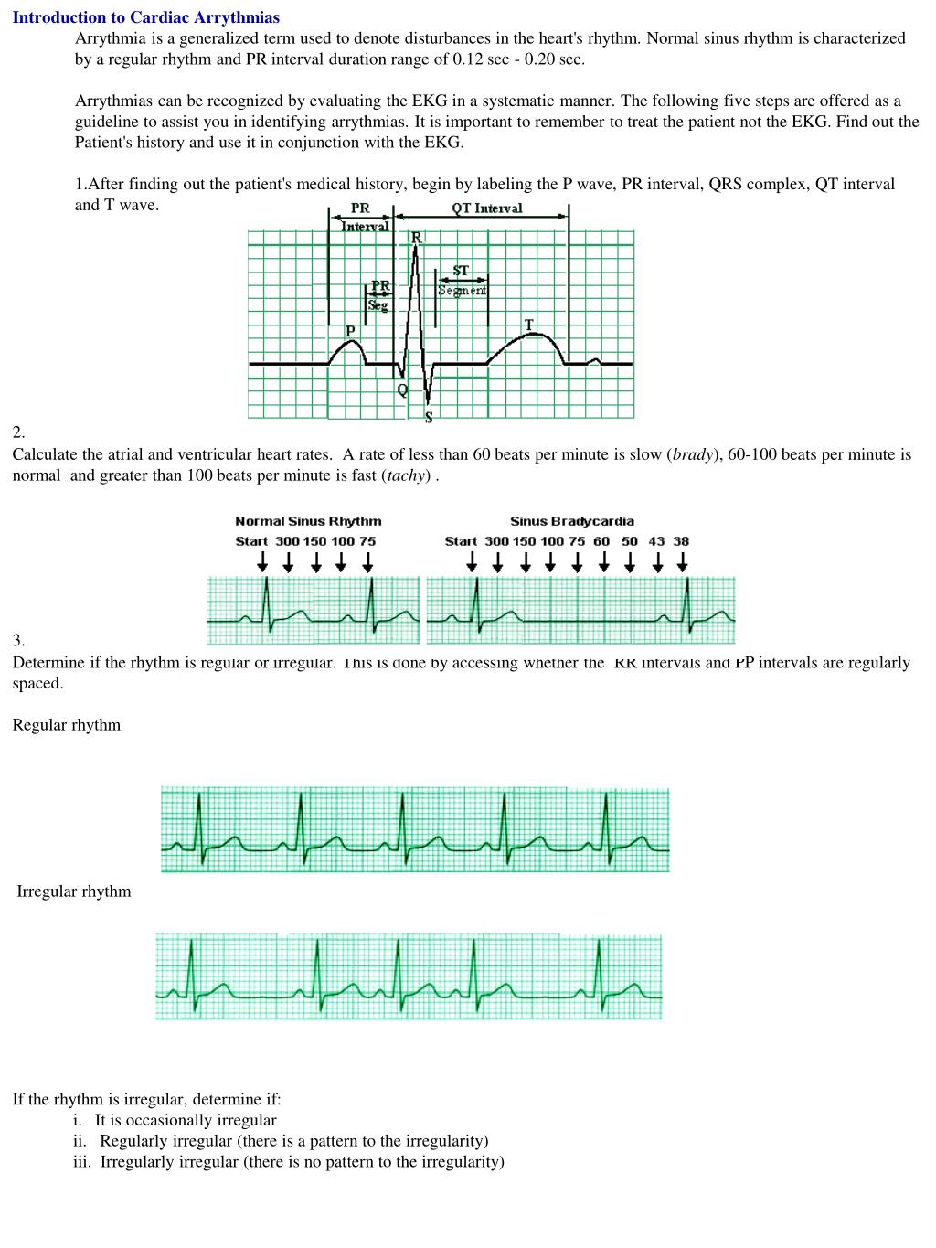
Introduction to Cardiac Arrythmias Arrythmia is a generalized term used to denote disturbances in the heart's rhythm. Normal sinus rhythm is characterized by a regular rhythm and PR interval duration range of 0.12 sec - 0.20 sec .
Introduction to Cardiac Arrythmias - slide 1
01 / 2Introduction to Cardiac Arrythmias
1 / 17

Télécharger la présentation

Introduction to Cardiac Arrythmias
Une image / un lien ci-dessous est fourni (tel quel) pour télécharger la présentation

Politique de téléchargement : le contenu du site vous est fourni TEL QUEL pour votre information et votre usage personnel ; il ne peut être vendu, concédé sous licence ni partagé sur d’autres sites sans l’accord de son auteur.Le contenu vous est fourni TEL QUEL, pour votre information et votre usage personnel uniquement. Téléchargez la présentation en cliquant sur ce lien. Lors du téléchargement, si vous ne parvenez pas à télécharger une présentation, l’éditeur a peut-être supprimé le fichier de son serveur.Pendant le téléchargement, si vous ne parvenez pas à obtenir une présentation, le fichier a peut-être été supprimé par l’éditeur.

F I N

Plus de contenu associé

Transcription de la présentation
  1. Introduction to Cardiac Arrythmias • Arrythmia is a generalized term used to denote disturbances in the heart's rhythm. Normal sinus rhythm is characterized by a regular rhythm and PR interval duration range of 0.12 sec - 0.20 sec. • Arrythmias can be recognized by evaluating the EKG in a systematic manner. The following five steps are offered as a guideline to assist you in identifying arrythmias. It is important to remember to treat the patient not the EKG. Find out the Patient's history and use it in conjunction with the EKG.  • 1.After finding out the patient's medical history, begin by labeling the P wave, PR interval, QRS complex, QT interval and T wave. • 2. • Calculate the atrial and ventricular heart rates.  A rate of less than 60 beats per minute is slow (brady), 60-100 beats per minute is normal  and greater than 100 beats per minute is fast (tachy) . • 3. • Determine if the rhythm is regular or irregular. This is done by accessing whether the  RR intervals and PP intervals are regularly spaced.  • Regular rhythm • Irregular rhythm • If the rhythm is irregular, determine if: •               i.   It is occasionally irregular •               ii.   Regularly irregular (there is a pattern to the irregularity) •               iii.  Irregularly irregular (there is no pattern to the irregularity)

  2. 4. Evaluate the waveform of the EKG in detail for additional clues: a. Determine the shape of the P-wave. Normally, P-waves are upright and each P wave is related to a QRS complex. If inverted, the impulse is  spreading from the ventricles to the atria in a retrograde manner.b. Determine if the PR interval is of normal length (0.12-0.2 seconds).c. Examine the QRS complexes and determine if the QRS complex is wide or narrow. Narrow QRS complexes (less than 0.12 sec) indicate that the rhythm is supraventricular (originating from above the ventricles). Wide QRS complexes (longer than 0.12 sec), indicate that the rhythm is originating in the ventricles or that there is an intraventicular block.d. Determine if the ST segment is displaced from the mid-line. 5. Review the patient's medical history, assess the patient to ensure that the assessment and rhythm agree. The following EKG clues can be used to recognize cardiac arrythmias in non sinus rhythm EKGs. If the rhythm is regular but too fast or slow, it could be an indication of either: a. Sinus bradycardia: The rhythm is regular and looks normal but is slower than 60 beats per minute. The RR interval is longer, often more than one second. P waves are present and regular and each P-wave is followed by a QRS complex in a ratio of 1:1. b. Sinus tachycardia: The  rhythm is regular and looks normal but at a rate greater than 100 beats per minute  The R-R interval is shorter (usually less than 0.6 seconds). P waves are present and regular and each P-wave is followed by a QRS complex in a ratio of 1:1. If the rhythm is irregular, check for the following: a. Atrial flutter. Atrial flutter waves (F-waves) with a characteristic saw-tooth form will also be observed at a rate of 200-350 BPM. b. Atrial fibrillation. No P-waves will observable. Rather, a wavy base-line is recorded. If there are no P-waves, it could be an indication of either: Sinus arrest with junctional or ventricular escape. If P-waves are not associated with QRS complexes, it could indicate either: 1. Ventricular tachycardia. 2. Third degree AV block.