Limb Repair and Regeneration
340 likes | 584 Vues
Limb Repair and Regeneration. Ricardo Gutierrez Biology 680 February 25, 2008. -Morphallaxis- lost body parts are replaced by remodeling of remaining part. -Occurs in Planarians -Epimorphosis- requires active cell proliferation before replacement

Limb Repair and Regeneration
E N D
Presentation Transcript
Limb Repair and Regeneration Ricardo Gutierrez Biology 680 February 25, 2008
-Morphallaxis- lost body parts are replaced by remodeling of remaining part. -Occurs in Planarians -Epimorphosis- requires active cell proliferation before replacement -Occurs in limb and fin regeneration, urodele amphibian and teleost fish
Epimorphosis characterized by formation of a blastema after wound healing http://www.eb.tuebingen.mpg.de/departments/3-genetics/zebrafish/christopher-antos/regeneration Zebrafish caudal fin after wound healing Bl=Blastema, We=Wound epidermis
Blastema is a mass of undifferentiated cells capable of growth and regeneration resulting from dedifferentiation of cells.
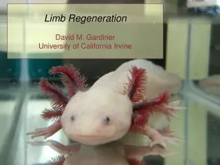
Zebrafish used as model organism for studies regeneration Most studies done on caudal fin -more accessible for surgery and manipulations
Fin regeneration divided into four successive steps • Wound healing • Blastema formation • Regenerative outgrowth concomitant to differentiation • Patterning of blastema http://www.eb.tuebingen.mpg.de/departments/3-genetics/zebrafish/christopher-antos/regeneration
Fins consist of two distinct ontogenic origins • Endoskeleton derived from endochondral bone located at base of fin • Exoskeleton derived from dermal origin located in the external part of the fin Regeneration is limited to the exoskeleton. Bone of each ray is called lepidotrichia Nature445, 311-314
Wound healing • Wound closure results from rapid apical migration of epithelial cells. • A dense mass of cells called the wound epidermis or apical epidermal cap form within 12 to 18 hours. • Migration of epithelial cells occur from close to the wound to several segments proximal to the wound. • There is no cellular proliferation
Two distinct layers are formed : -An outer layer with compact flat cells -Basal layer with cuboidal cells. -The rest of the cells form a loose multilayered mass of cells between the two distinct layers Cytoplasmic protrusions occur from the basal epithelial layer into the underlying mesenchymal cells. Geraudie and singer showed that exchange between epithelial and mesenchymal cells necessary for blastema formation
Poss et al showed that wnt 5 and lef 1 are activated by epithelial cells lining the mesenchymal cells Other studies have shown that epithelial-mesenchymal interactions are required for organ formation.
Balstema formation In urodele amphibians dedifferentiation of firbroblast-like cells occurs leading to proliferation. There is also proliferation of Scleroblasts, which line the hemirays and endothelial cells. -Scleroblast play a role in bone repair -Endothelial cells are involved in revascularization of he fin.
It has also been proposed that stem cells could lead provide cells for the formation of the blastema along with the dedifferentiation of cells. The only stem cells that have shown to develop in zebrafish are melanocytes from unpigmented precursor. Mesenchymal cells also migrate from distant locations and become part of the blastema. • Akimenko and Smith conclude that there are two cellular mechanisms: • Cell dedifferentiation • Activation of reserve cells.
FGF Fibroblast growth factor (FGF) plays a role in amphibian limb regeneration. FGF signaling has been shown to be necessary in blastema formation Poss et al. (2000b) used SU 5402, an FGFr1 inhibitor, following amputation. -SU 5402 prevented blastema formation , but did not affect wound healing. -SU 5402 did not prevent disorganized aspect of cells where the blastema would have formed.
Msx Msx homeobox gene family also involved in epithilial-mesenchymal interactions Msx products play a role in organogenesis Han et al. (2003) demonstrated that msx is required for distal digit regeneration in fetal mise. Msx proteins are used in signaling pathways involving Bmp, Fgf and SHH During blastema formation msxa and msxd transcripts restricted to the epithelial cap -msxb and msxc are localized in proliferating cells in the blastema SU5402 also inhibit expression of msxb and msxc Han et al. (2003)
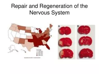
-Studies show that the nervous system also plays a critical role in fin and limb regeneration -resection of the nerves in the brachial plexus prevents blastema formation and outgrowth, but does not affect wound healing. -Factors involved with regeneration and the nervous system are still unknown.
In urodele, medium to late blastema buds do not require nerves for limb formation, but are required for the early blastema formation. http://www.mun.ca/biology/desmid/brian/BIOL3530/DB_Ch13/DBNRegen.html
Outgrowth of the regenerate During regenerative outgrowth the blastema becomes divided into two populations with different cell cycles -Proximal blastema (PB) cells -proliferate at a faster rate than during blastema formation -Distalmost blastema (DB) -very low cycling to a nonproliferation rate.
Only PB cells express proliferating cell nuclear antigen (PCNA). Proliferation is associated with modifications of msxb expression. Msxb becomes restricted to DB during outgrowth Suggested that msxb-expressing cells establish a boundary providing direction for regenerative outgrowth
Poss et al. demonstrated that mps1 are coexpressed with msxb in the blastema -During regenerative outgrowth mps1 and msxb segregate. -msxb in the DB, poor proliferation -mps1 in PB, high poliferation -This suggests that mps1 required to maintain high proliferation in the PB.
Differentiation and pattern Formation Differentiation starts while the blastema continues the outgrowth phase. -Differentiation starts with cells located at the proximal part of the PB. -Blastema cells lining the epithelial tissue and located between the PB and the patterning zone differentiate into scleroblasts. -Scleroblast will secrete the bone matrix forming the lepidotrichia.
-Bone matrix will mineralize in a proximal-distal direction -the bones regenerate by formation of new segments. -Scleroblasts at the joint express evx1, suggesting that evx1 involved in joint formation. -expression of hoxa11 and hoxa13b in scleroblasts suggests that these factors are involved in bone cell differentiation.
Tetrapod limb regeneration Regeneration can be broken into a sequence of stages from amputation to a fully developed regenerative limb Time need varies with urodele specie and size of organism Dr. Susan Bryant, Univ. of Calif., Irvine
As with zebrafish after amputation epidermis migrates to cover the wound (WE) and an apical epidermal cap (AEC) forms. Formation of the blastema is dependent on factors produced by the WE and AEC. Regeneration progresses from proximal to distal and from anterior to posterior.
All tetrapods regenerate at some stage in the life history of the organism. Regenerative ability is intrinsic property of limb cells and not hormonal or other changes in animals In studies young limb buds grafted to regenative-incompetent older host regenerate, whereas older limb buds grafted to young regenative-competent host are unable to regenerate.
Loss of regenerative ability in Xenopus is associated with inability reactivate expression of genes involved in growth and patter formation, such as Shh. Treatment of regeneration-incompetent Xenopus limbs with Fgf partially rescues regenerative ability. In mammals the ability to regenerate the most distal structures, terminal phalanx digits, is retained. If the wound is sutured the regenerative response is inhibited. Han et al. (2003)
Regeneration requires two steps. First step involves formation of the blastema, preparation phase I. Second step involves the redevelopment of the limb, redevelopment phase II.
Stages of development are comparable to those of regeneration, during the phase II. Both hoxa9 and hoxa13 are expressed on in the autopod, while only hoxa9 is expressed in the zeugopod. Hoxa9 diagonal, hoxa13 and hoxa9 black Phase I gene expression is unique to regeneration Phase I Phase II
Cell contributions to blastema formation -Nerves and blood vessels are formed by the structures that are left behind do to amputation. -As mentioned before a nerve supply is required for regeneration to occur. -Skeletal tissues have a minor role in contributing to the blastema. -early studies concluded that periosteum and perichondrium contributed to the blastema
Muscle tissue becomes mononucleated in the blastema through an unknown mechanism. -the cells contribute mostly to regenerating muscle, but some form cartilage This suggests that there is some transdifferentiation.
Connective tissue fibroblasts have the greatest contribution in number of cells and control of growth and pattern formation. On average fibroblast contribute about 42 % of cell mass in the blastema. Fibroblast tissues are the only mature limb tissues the influence pattern formation determining final regenerate orientation.
Positional information is demonstrated by induction of growth and pattern formation. A) Control. B) Graft from the left hindlimb bud was placed onto a right regenerating hindlimb stump
http://8e.devbio.com/printer.php?ch=1&id=184 -Results of grafting blastema cells from anterior and posterior positions within the limb demonstrated that cells from different limb positions express different cell surface molecules -If cells express similar cell surface molecules there is no outgrowth.
Fibroblast cells show high degree of development and physiological plasticity, they are basically blastema cells. -have ability to give rise to many cell types. -Studies of bone induction using BMP target fibroblast that are responsive to BMP signaling. -It has not been presumed that fibroblasts go through dedifferentiation then transdifferentiation into chondrocytes or osteocytes.
Reference Han, M et al. (2003) Digit regeneration is regulated by Msx1 and BMP4 in fetal mice.Development 130, 5123-5132