Download
1 / 45
510 likes | 973 Vues
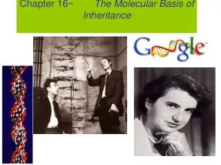
Chapter 16~ The Molecular Basis of Inheritance. Scientific History . The march to understanding that DNA is the genetic material T.H. Morgan (1908) Frederick Griffith (1928) Avery, McCarty & MacLeod (1944) Erwin Chargaff (1947) Hershey & Chase (1952) Watson & Crick (1953)

E N D
Scientific History • The march to understanding that DNA is the genetic material • T.H. Morgan (1908) • Frederick Griffith (1928) • Avery, McCarty & MacLeod (1944) • Erwin Chargaff (1947) • Hershey & Chase (1952) • Watson & Crick (1953) • Meselson & Stahl (1958)
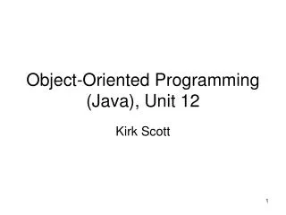
The “Transforming Principle” 1928 • Frederick Griffith • Streptococcus pneumonia bacteria • was working to find cure for pneumonia • harmless live bacteria (“rough”) mixed with heat-killed pathogenic bacteria (“smooth”) causes fatal disease in mice • a substance passed from dead bacteria to live bacteria to change their phenotype • “Transforming Principle”
The “Transforming Principle” mix heat-killed pathogenic & non-pathogenic bacteria live pathogenic strain of bacteria live non-pathogenic strain of bacteria heat-killed pathogenicbacteria A. B. D. C. mice die mice live mice live mice die Transformation=change in phenotype something in heat-killed bacteria could still transmit disease-causing properties
DNA is the “Transforming Principle” 1944 • Avery, McCarty & MacLeod • purified both DNA & proteins separately from Streptococcus pneumonia bacteria • which will transform non-pathogenic bacteria? • injected protein into bacteria • no effect • injected DNA into bacteria • transformed harmless bacteria into virulent bacteria mice die What’s the conclusion?
Avery, McCarty & MacLeod 1944 | ??!! • Conclusion • first experimental evidence that DNA was the genetic material Oswald Avery Maclyn McCarty Colin MacLeod
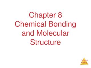
Confirmation of DNA 1952 | 1969 Hershey • Hershey & Chase • classic “blender” experiment • worked with bacteriophage • viruses that infect bacteria • grew phage viruses in 2 media, radioactively labeled with either • 35S in their proteins • 32P in their DNA • infected bacteria with labeled phages Why useSulfurvs.Phosphorus?
Hershey & Chase Protein coat labeled with 35S DNA labeled with 32P T2 bacteriophages are labeled with radioactive isotopes S vs. P bacteriophages infect bacterial cells bacterial cells are agitated to remove viral protein coats Which radioactive marker is found inside the cell? Which molecule carries viral genetic info? 32P radioactivity foundin the bacterial cells 35S radioactivity found in the medium
Blender experiment • Radioactive phage & bacteria in blender • 35S phage • radioactive proteins stayed in supernatant • therefore viral protein did NOT enter bacteria • 32P phage • radioactive DNA stayed in pellet • therefore viral DNA did enter bacteria • Confirmed DNA is “transforming factor” Taaa-Daaa!
Hershey & Chase 1952 | 1969 Hershey Martha Chase Alfred Hershey
Chargaff 1947 • DNA composition: “Chargaff’s rules” • varies from species to species • all 4 bases not in equal quantity • bases present in characteristic ratio • humans: A = 30.9% T = 29.4% G = 19.9% C = 19.8% RulesA = T C = G That’s interesting!What do you notice?
Structure of DNA 1953 | 1962 • Watson & Crick • developed double helix model of DNA • other leading scientists working on question: • Rosalind Franklin • Maurice Wilkins • Linus Pauling Wilkins Pauling Franklin
1953 article in Nature Watson and Crick Watson Crick
Double helix structure of DNA “It has not escaped our notice that the specific pairing we have postulated immediately suggests a possible copying mechanism for the genetic material.” Watson & Crick
Directionality of DNA nucleotide • You need to number the carbons! • it matters! PO4 N base 5 CH2 This will beIMPORTANT!! O 1 4 ribose 3 2 OH
The DNA backbone 5 PO4 • Putting the DNA backbone together • refer to the 3 and 5 ends of the DNA • the last trailing carbon base CH2 5 O 4 1 C 3 2 O P –O O Sounds trivial, but…this will beIMPORTANT!! O base CH2 5 O 4 1 2 3 OH 3
Anti-parallel strands • Nucleotides in DNA backbone are bonded from phosphate to sugar between 3 & 5 carbons • DNA molecule has “direction” • complementary strand runs in opposite direction 5 3 3 5
hydrogen bonds covalent phosphodiester bonds Bonding in DNA 5 3 3 5 ….strong or weak bonds? How do the bonds fit the mechanism for copying DNA?
Base pairing in DNA • Purines • adenine (A) • guanine (G) • Pyrimidines • thymine (T) • cytosine (C) • Pairing • A : T • 2 bonds • C : G • 3 bonds
But how is DNA copied? • Replication of DNA • base pairing suggests that it will allow each side to serve as a template for a new strand “It has not escaped our notice that the specific pairing we have postulated immediately suggests a possible copying mechanism for the genetic material.” —Watson & Crick
Copying DNA • Replication of DNA • base pairing allows each strand to serve as a template for a new strand • new strand is 1/2 parent template & 1/2 new DNA • semi-conservativecopy process
Semiconservative replication, • when a double helix replicates each of the daughter molecules will have one old strand and one newly made strand. • Experiments in the late 1950s by Matthew Meselson and Franklin Stahl supported the semiconservative model, proposed by Watson and Crick, over the other two models. (Conservative & dispersive)
DNA Replication Let’s meetthe team… • Large team of enzymes coordinates replication
Replication: 1st step • Unwind DNA • helicase enzyme • unwinds part of DNA helix • stabilized by single-stranded binding proteins helicase single-stranded binding proteins replication fork
Replication: 2nd step • Build daughter DNA strand • add new complementary bases • DNA polymerase III DNA Polymerase III
Replication 3 5 energy DNA Polymerase III • Adding bases • can only add nucleotides to 3 end of a growing DNA strand • need a “starter” nucleotide to bond to • strand only grows 53 DNA Polymerase III energy DNA Polymerase III energy DNA Polymerase III energy 3 5
Okazaki ligase 3 3 3 3 3 3 3 5 5 5 5 5 5 5 Leading & Lagging strands Limits of DNA polymerase III • can only build onto 3 end of an existing DNA strand Okazaki fragments Lagging strand growing replication fork Leading strand Lagging strand • Okazaki fragments • joined by ligase • “spot welder” enzyme DNA polymerase III Leading strand • continuous synthesis
DNA polymerase III 3 3 3 3 3 3 3 3 3 3 3 growing replication fork growing replication fork 5 5 5 5 5 5 5 5 5 5 5 5 5 5 5 5 Replication fork / Replication bubble leading strand lagging strand leading strand lagging strand leading strand lagging strand
3 3 3 3 3 3 DNA polymerase III 5 5 5 5 5 5 Starting DNA synthesis: RNA primers Limits of DNA polymerase III • can only build onto 3 end of an existing DNA strand growing replication fork primase RNA RNA primer • built by primase • serves as starter sequence for DNA polymerase III
3 3 3 3 3 3 DNA polymerase III 5 5 5 5 5 5 Starting DNA synthesis: RNA primers Limits of DNA polymerase III • can only build onto 3 end of an existing DNA strand growing replication fork primase RNA RNA primer • built by primase • serves as starter sequence for DNA polymerase III
ligase 3 3 3 3 5 5 5 5 Replacing RNA primers with DNA DNA polymerase I • removes sections of RNA primer and replaces with DNA nucleotides DNA polymerase I growing replication fork RNA But DNA polymerase I still can only build onto 3 end of an existing DNA strand
3 3 3 3 5 5 5 5 Houston, we have a problem! Chromosome erosion All DNA polymerases can only add to 3 end of an existing DNA strand DNA polymerase I growing replication fork DNA polymerase III RNA Loss of bases at 5 endsin every replication • chromosomes get shorter with each replication • limit to number of cell divisions?
3 3 3 3 5 5 5 5 Telomeres Repeating, non-coding sequences at the end of chromosomes = protective cap • limit to ~50 cell divisions growing replication fork telomerase Telomerase • enzyme extends telomeres • can add DNA bases at 5 end • different level of activity in different cells • high in stem cells & cancers -- Why? TTAAGGG TTAAGGG TTAAGGG
direction of replication Replication fork DNA polymerase III lagging strand DNA polymerase I 3’ primase Okazaki fragments 5’ 5’ ligase SSB 3’ 5’ 3’ helicase DNA polymerase III 5’ leading strand 3’ SSB = single-stranded binding proteins
Roger Kornberg 2006 Arthur Kornberg 1959 DNA polymerases • DNA polymerase III • 1000 bases/second! • main DNA builder • DNA polymerase I • 20 bases/second • editing, repair & primer removal DNA polymerase III enzyme
Editing & proofreading DNA • 1000 bases/second = lots of typos! • DNA polymerase I • proofreads & corrects typos • repairs mismatched bases • removes abnormal bases • repairs damage throughout life • reduces error rate from 1 in 10,000 to 1 in 100 million bases
Fast & accurate! • It takes E. coli <1 hour to copy 5 million base pairs in its single chromosome • divide to form 2 identical daughter cells • Human cell copies its 6 billion bases & divide into daughter cells in only few hours • remarkably accurate • only ~1 error per 100 million bases • ~30 errors per cell cycle
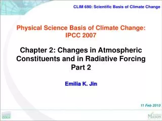
1 2 3 4 What does it really look like?
DNA Packing DNAdoublehelix(2-nmdiameter Histones “Beads ona string” Nucleosome(10-nm diameter) Tight helical fiber(30-nm diameter) Supercoil(200-nm diameter) 700nm Metaphase chromosome
Nucleosomes 8 histone molecules • “Beads on a string” • 1st level of DNA packing • histone proteins • 8 protein molecules • positively charged amino acids • bind tightly to negatively charged DNA
DNA packing as gene control • Degree of packing of DNA regulates transcription • tightly wrapped around histones • no transcription • genes turned off • heterochromatin darker DNA (H) = tightly packed • euchromatin lighter DNA (E) = loosely packed H E