THE AUTONOMIC NERVOUS SYSTEM
530 likes | 552 Vues
Explore the functions, structures, and differences between the somatic and autonomic nervous systems. Learn about motor neurons, ganglia, neurotransmitters, and the sympathetic and parasympathetic divisions.

THE AUTONOMIC NERVOUS SYSTEM
E N D
Presentation Transcript
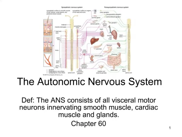
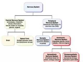
THE AUTONOMIC NERVOUS SYSTEM • System of motor neurons that innervates smooth and cardiac muscle and glands • Also called: • The involuntary nervous system,which reflects its subconscious control • The general visceral motor system, which indicates the location of most of its effectors
COMPARISON OF THE SOMATIC AND AUTONOMIC NERVOUS SYSTEM • The somatic nervous system stimulates skeletal muscles, while the ANS innervates cardiac and smooth muscle and glands
COMPARISON OF THE SOMATIC AND AUTONOMIC NERVOUS SYSTEM • Ganglion: collection of nerve cell bodies outside the CNS • Somatic Motor Division: lacks ganglia entirely • The dorsal root ganglia are part of the sensory, not the motor, division of the peripheral nervous system • In the somatic nervous system, the cell bodies of the neurons are in the spinal cordand their axons extend to the skeletal muscles they innervate • Cell bodies of the motor neurons are in the CNS, and their axons extend in spinal nerves all the way to the skeletal muscles they serve • Somatic motor fibers are typically thick, heavily myelinated fibers that conduct nerve impulses very rapidly
COMPARISON OF THE SOMATIC AND AUTONOMIC NERVOUS SYSTEM • Motor unit of the ANS is a two-neuron chain: • The cell body of the first neuron, the preganglionic neuron, resides in the brain or spinal cord: • Its axon, called the preganglionic axon, synapses with the second motor neuron, the ganglionic neuron, in an autonomic ganglion outside the CNS • The axon of the ganglionic neuron, called the postganglionic axon, then extends to the effector organ • Ganglia are motor ganglia, containing the cell bodies of motor neurons • Preganglionic axons are lightly myelinated, thin fibers • Postganglionic axons are even thinner and are unmyelinated • Conduction through the autonomic efferent chain is slower than conduction in the somatic motor chain
SOMATIC VS AUTONOMIC • Somatic Division: • Axons of somatic motor neurons extend from the CNS to their effectors (skeletal muscle cells) • These axons are typically heavily myelinated • Somatic motor neurons release acetylcholine, and the effect is always stimulatory
SOMATIC VS AUTONOMIC • Autonomic Division: • Axons of most preganglionic neurons run from the CNS to synapse in a peripheral autonomic ganglion with a ganglionic neuron • A few sympathetic preganglionic axons synapse with cells of the adrenal medulla
SOMATIC VS AUTONOMIC • Autonomic Division: • Postganglionic axons run from the ganglion to the effectors (cardiac and smooth muscle fibers and glands) • Preganglionic axons are lightly myelinated • Postganglionic axons are unmyelinated
SOMATIC VS AUTONOMIC • Autonomic Division: • All preganglionic fibers release acetylcholine • All parasympathetic postganglionic fibers release acetylcholine • Most sympathetic postganglionic fibers release norepinephrine • Stimulated adrenal medullary cells release norepinephrine and epinephrine into the blood
SOMATIC VS AUTONOMIC • Autonomic Division: • Autonomic effects are stimulatory or inhibitory, depending on the postganglionic neurotransmitter released and the receptor types on the effectors
COMPARISON OF THE SOMATIC AND AUTONOMIC NERVOUS SYSTEM • The neurotransmitter released by the somatic motor neurons is acetylcholine, which always has an excitatory effect • The neurotransmitters released by the ANS are epinephrine and acetylcholine, and both may have either an excitatory or an inhibitory effect • There is overlap between the somatic and autonomic nervous systems, and most body responses to changing internal and external stimuli involve both skeletal muscle activity and visceral organ responses
DIVISIONS OF THE AUTONOMIC NERVOUS SYSTEM • The parasympathetic division keeps body energy use as low as possible while directing digestion and elimination activities (most active in nonstressful situations) • Relax after a meal • The sympathetic division prepares the body to respond to an emergency or threatening situation • Fight or flight system • Visceral (and perhaps cutaneous) blood vessels are constricted, and blood is shunted to active skeletal muscles and heart • Increased ventilation (dilation of bronchioles) • Increase sugar release by liver into blood
PARASYMPATHETIC/SYMPATHETIC • Think of the parasympathetic division as the D: • Digesting, defecation, diuresis (urination) • Think of the sympathetic division as the E: • Exercise, excitement, emergency, and embarrassment • A dynamic antagonism exists between the two: fine adjustments are made continuously by both
PARASYMPATHETIC (CRANIOSACRAL) DIVISION • The preganglionic axons extend from the CNS nearly all the way to the structures to be innervated where they synapse with ganglionic neurons in the terminal ganglia • The cranial outflow consists of preganglionic fibers that run in the oculomotor, facial, glossopharyngeal, and vagus cranial nerves • The rest of the large intestine and the pelvic organs are served by the sacral outflow, which arises from neurons located in the lateral gray matter of spinal cord segments
PARASYMPATHETIC (CRANIOSACRAL) DIVISION • Origin sites: • Parasympathetic fibers emerge from the brain and sacral spinal cord • Sympathetic fibers originate from the thoracolumbar region of the spinal cord
PARASYMPATHETIC (CRANIOSACRAL) DIVISION • Different lengths of their fibers: • Parasympathetic: long preganglionic and short postganglionic fibers • Sympathetic: short preganglionic and long postganglionic fibers
PARASYMPATHETIC (CRANIOSACRAL) DIVISION • Location of ganglia: • Parasympathetic: • Most ganglia are located in the visceral effector organs • Sympathetic: • Ganglia lie close to the spinal cord
PARASYMPATHETIC (CRANIOSACRAL) DIVISION of the ANS • Solid lines indicate preganglionic nerve fibers • Dashed lines indicate postganglionic fibers • Terminal ganglia of the vagus and pelvic splanchnic nerve fibers are not shown • Most of these ganglia are located in or on the target organ
PARASYMPATHETIC (CRANIOSACRAL) DIVISION of the ANS • Cranial: • Oculomotor Nerves (III): • Innervates smooth muscles of the eye • Facial Nerves (VII): • Nasal/Lacrimal Glands • Submandibular/Sublingual Glands • Glossopharyngeal Nerves (IX): • Parotid Salivary Gland • Vagus Nerves (X): • Serves virtually every organ in the thoracic and abdominal cavities • Sacral: • Pelvic Nerves (S2+S4): • Distal half of large intestine, urinary bladder, ureters, and reproductive organs
SYMPATHETIC(THORACOLUMBAR)DIVISION • The sympathetic division supplies the visceral organs in the internal body cavities but also all visceral structures in the somatic part of the body • When synapses are made in chain ganglia, the postganglionic axons enter the ventral (or dorsal) ramus of the adjoining spinal nerves by way of communicating branches called gray rami communicates • The preganglionic fibers from T5 down synapse in collateral ganglia; thus these fibers enter and leave the sympathetic chains without synapsing • Some fibers of the thoracic splanchnic nerves terminate by synapsing with the hormone producing medullary cells of the adrenal cortex
SYMPATHETIC(THORACOLUMBAR)DIVISION • The sympathetic division supplies the visceral organs in the internal body cavities but also all visceral structures in the somatic part of the body • When synapses are made in chain ganglia, the postganglionic axons enter the ventral (or dorsal) ramus of the adjoining spinal nerves by way of communicating branches called gray rami communicates • The preganglionic fibers from T5 down synapse in collateral ganglia; thus these fibers enter and leave the sympathetic chains without synapsing • Some fibers of the thoracic splanchnic nerves terminate by synapsing with the hormone producing medullary cells of the adrenal cortex
SYMPATHETIC(THORACOLUMBAR)DIVISION • Head: • Inhibits nasal and salivary glands • Reason our mouth goes dry when scared • Thorax: • Heart • Thyroid gland • Skin • Lungs • Esophagus • Abdomen: • Stomach • Intestines (except distal region) • Liver • Spleen • Kidneys • Pelvis: for the most part, the activities of these organs is inhibited • Distal half of large intestine • Urinary bladder • Reproductive organs
SYMPATHETIC (THORACOLUMBAR) DIVISIONof theANS • Solid lines:indicate preganglionic fibers • Dashed lines:indicate postganglionic fibers • The least thoracic and sacral splanchnic nerves are not shown
SYMPATHETIC TRUNKS and PATHWAYS • (a): The organs of the anterior thorax have been removed to allow visualization of the chain ganglia of the sympathetic trunks
SYMPATHETIC TRUNKS and PATHWAYS • (b): Three sympathetic pathways: • 1. Synapse in a chain (paravertebral) ganglion at same level • 2. Synapse in a chain ganglion at a different level • 3. Synapse in a collateral (prevertebral) ganglion anterior to the vertebral column
The visceral sensory neurons are thefirst link in autonomic reflexes by sending information concerning chemical changes, stretch, and irritation of the viscera
VISCERAL REFLEXES • Visceral reflex arcs have essentially the same components as somatic reflex arcs: receptor, sensory neuron, integration center, motor neuron, and effector • Except: a visceral reflex arc has a two-neuron motor chain
VISCERAL REFLEXES • Visceral reflexes have the same elements as somatic reflexes but occur over polysynaptic pathways because of the two-motor-neuron efferent pathway • The integration center may involve a dorsal horn interneuron, or it may just be a synapse with a preganglionic neuron (as shown) • The visceral afferent fibers are found both in spinal nerves (as depicted here) and in autonomic nerves
REFERRED PAIN • Anterior cutaneous areas to which pain from certain visceral organs is referred • The fact that visceral pain afferents travel along the same pathways as somatic pain fibers explains this phenomenon • Pain stimuli arising in the viscera are perceived as somatic in origin: • E.g.: a heart attack may produce a sensation of pain that radiates to the superior thoracic wall and along the medial aspect of the left arm
ANS PHYSIOLOGY • Neurotransmitters: • Acetylcholine (ACh) and norepinephrine (NE) (originally called noradrenaline) are the major neurotransmitters released by ANS neurons • Acetylcholine secreted by cholinergic fibers: • Somatic motor neurons • All preganglionic axons of ANS • All parasympathetic postganglionic axons at synapses with their effectors • Releasing fibers are called cholinergic • Cholinergic receptors, such as nicotinic and muscarinic receptors, bind acetylcholine
ANS PHYSIOLOGY • Neurotransmitters: • Most sympathetic postganglionic axons release norepinephrine (NE) • Classified as adrenergic fibers (fibers that release epinephrine (adrenaline) or norepinephrine (noradrenaline) • Only exception: fibers innervating the sweat glands of the skin, some blood vessels within skeletal muscles and the external genitalia, all of which secrete Ach
ANS PHYSIOLOGY • Norepinephrine (Noradrenaline): • A hormone produced by the Adrenal Medulla • A catecholamine (biologically active amine derived from the amino acid tyrosine) • Similar in chemical and pharmacological properties to epinephrine • Vasoconstrictor with little effect on cardiac output • Effect on raising blood pressure • Neurotransmitter • Epinephrine (Adrenaline): • A hormone produced by the Adrenal Medulla • A catecholamine (biologically active amine derived from the amino acid tyrosine) • Produced by the adrenal gland • Maintains blood pressure and cardiac output • Keeps airways open (dilates bronchioles) • Increases metabolic activities • Raises blood sugar level
ANS PHYSIOLOGY • The effects of ACh and NE on their effectors are not consistently either excitation or inhibition • Response of visceral effectors to these neurotransmitters depends not onlyon the neurotransmitters but also on the receptors to which they attach
ANS PHYSIOLOGY • Cholinergic Receptors (Ach): • Nicotinic receptors: • Binds ACh and mimics it effects • Found on: • Motor end plates of skeletal muscles (somatic) • All ganglionic neurons (sympathetic and parasympathetic) • Hormone-producing cells of Adrenal Medulla • Effect of ACh binding to nicotinic receptors is alwaysstimulatory and results in excitation of the neuron or effector cell • Muscarine receptors: • A mushroom poison (muscarine) activates a different set of Ach receptors • Occur on all effector cells stimulated by postganglionic cholinergic fibers • all parasympathetic target organs and a few sympathetic targets such as eccrine sweat glands and some blood vessels of skeletal muscles • Effect of ACh binding is inhibitory or stimulatory depending on the target organ • E.g.: slows heart rate / increases smooth muscle contraction in gastrointestinal tract
ANS PHYSIOLOGY • Adrenergic Receptors: • Two major classes: • alpha and beta • In general: • NE (or epinephrine) binding to a alpha receptor is stimulatory • NE (or epinephrine) binding to a beta receptor is inhibitory • Exception: increase heart activity ( reflects the fact that there are subclasses)
ANS PHYSIOLOGY • Knowing the locations of the cholinergic and adrenergic receptor subtypes allows specific drugs to be prescribed to obtain desired inhibitory or stimulatory effects on target organs • Atropine: prevents salivation / dries respiratory system secretions / dilates pupil • Counteracts the effects of parasympathetic stimulation • Anti-Depression drugs: prolong activity of NE on postsynaptic membrane • Block the reuptake of norepinephrine and serotonin • Colds / coughs / allergies • Beta blockers: heart and blood pressure • Inhibits the activities of the sympathetic nervous system and adrenergic hormones
ANS PHYSIOLOGY • Interactions of the autonomic divisions • The sympathetic division mediates reflexes that regulate body temperature, release renin (enzyme stimulating vasoconstriction) from the kidneys, and promote metabolic effects • The parasympathetic division exerts short-lived, localized control of its effectors, while the sympathetic division responds in a diffuse and interconnected way to cause a body-wide mobilization
ANS PHYSIOLOGY • Interactions of the autonomic divisions: • Most visceral organs receive dual innervations by both ANS divisions, allowing for a dynamic antagonism to exist between the divisions and precise control of visceral activity • Sympathetic division will increase heart and respiratory rates (dilate bronchioles) during a fight-or-flight situation and the parasympathetic can slow the heart and respiratory rates (constrict bronchioles) • Parasympathetic division increase activities of digestive muscles (peristalsis)and elimination organs (relaxes anal sphincters) while the sympathetic division decreases the peristalsis and constricts anal sphincters • Sympathetic tone (slight muscle contraction) occurs in the vascular system, and parasympathetic tone (slight muscle contraction) occurs in the digestive and urinary tracts
ANS PHYSIOLOGY • Interactions of the autonomic divisions • The parasympathetic and sympathetic divisions may work together to achieve a common purpose • For example: • In the male, the parasympathetic division controls erection (vasodilation) of the penis while the sympathetic division controls ejaculation • In the female, the parasympathetic division controls erection (vasodilation) of the clitoris while the sympathetic division controls reverse peristalsis in the vagina • Explains why sexual performance is sometimes impaired when people are anxious or upset and the sympathetic division is in charge
ANS PHYSIOLOGY • Control of Autonomic Functioning: • The brain stem appears to exert the most direct influence over autonomic functions • The hypothalamus is the main integration center for the autonomic nervous system • Cortical or voluntary control of the autonomic nervous system does appear to be possible