Download
1 / 20
200 likes | 516 Vues
Academic Half-Day The Chemical Basis for Neuronal Communication. Marie-Pierre Thibeault-Eybalin, R4 November 5 th , 2008. Introduction. 100 billion (10 11 ) neurons in the brain Up to 100,000 terminal contacts / neuron 10 16 connections between neurons / brain Connections = Synapses

E N D
Academic Half-DayThe Chemical Basis for Neuronal Communication Marie-Pierre Thibeault-Eybalin, R4 November 5th, 2008
Introduction • 100 billion (1011) neurons in the brain • Up to 100,000 terminal contacts / neuron • 1016 connections between neurons / brain • Connections = Synapses • Chemical messenger is released at pre-synaptic membrane of axon or dendrite terminal • It travels across synaptic cleft • It binds onto its receptor on post-synaptic membrane of other neuron • It activates effector system • Chemical messenger may be released at non-synaptic locations to influence distant neurons
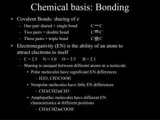
Criteria to define chemical messenger as neurotransmitter • Localization: A putative neurotransmitter must be localized to the presynaptic elements of an identified synapse and must be present also within the neuron from which the presynaptic terminal arises. • Release: The substance must be shown to be released from the presynaptic element upon activation of that terminal and simultaneously with depolarization of the parent neuron. • Identity: Application of the putative neurotransmitter to the target cells must be shown to produce the same effects as those produced by stimulation of the neurons in question.
Synaptic transmission • Variable synaptic delay from pre-synaptic neurotransmitter release to excitation or inhibition of post-synaptic neuron • Synaptic delay depends on complexity of transduction mechanisms at post-synaptic membrane
Regulatory mechanisms • To regulate amount of neurotransmitter release • Pre-synaptic receptor-mediated autoregulation • Neurotransmitter in synaptic cleft binds to pre-synaptic receptor • Inhibitory feedback mechanism • Retrograde transmission • 2nd chemical messenger diffuses from post-synaptic to pre-synaptic membranes, e.g. NO
Secretory vesicles • Small = Synaptic vesicles • For small molecules • Synthesized within vesicles, e.g. NE or uploaded by high-affinity ATP-proton-coupled transporters in terminals, e.g. ACh • Recycled • 50 nm diameter • Cluster in active zones • Large dense-cored vesicles • For neuropeptides, "built-in" in neuronal soma ± co-stored small molecule • Not-recycled • 75-150 nm diameter • Found in intraneuronal locations + terminals, less numerous • Neurosecretory vesicles • Hypothalamic neuron terminals in neurohypophysis • For neurohormones • 150-200 nm diameter
Exocytic release Fusion pore Docking complex
Signal transduction • Most receptors are transmembrane glycoproteins • Binding of neurotransmitter to receptor induces conformational change • 4 transduction mechanisms • Ligand-gated ion channels • G-protein-coupled receptors • Enzymes e.g. tyrosine kinase • Ligand-dependent regulators of nuclear transcription e.g. testosterone • Receptors often named after family of neurotransmitters they bind e.g. cholinergic and adrenergic receptors • Multiple subtypes based on response • Nicotinic ACh receptors usually excitatory • Muscarinic ACh receptors usually inhibitory • Individual neurotransmitter family members of have different potency • Rank order of potency according to EC50% • Concentration of individual neurotransmitter required to reach 50% of maximal response expected • The same neurotransmitter may have excitatory or inhibitory responses depending on receptor type
Structure of neurotransmitter receptors Ligand-gated ion channels • Multiple subunits = transmembrane glycoproteins connected via intra-and extra-cellular loops • Cylindrical • Binding site in transmembrane portion • Conformation changes opens gate inside channel • Selectively pass small ions • 2 genetic families based on AA sequence homology • Nicotinic ACh, serotonin, GABA, glycine • Glutamate
G-protein-coupled receptors • Glycoprotein chains with multiple transmembrane loops • -helices • β-pleated sheets • Binding site in transmembrane or extra-cellular portion • 3 components • Receptor • GTP-binding heterotrimer • Effector protein (enzyme or ion channel) • Examples • Rhodopsin • Odorants • Biogenic amines • Bioactive peptides • β2-adrenergic receptor
Receptor regulation • Desensitization • Reduction in receptor agonist-induced response after seconds to minutes of stimulation mediated by conformational changes • Homologous • Heterologous • Phosphorylation of intracellular portion of receptor altering its binding affinity • Downregulation of receptor number at post-synaptic membrane • Internalization of receptor by invagination of post-synaptic membrane
Maintenance of synaptic environment • To reduce or eliminate neurotransmitters in synaptic cleft • Enzymatic degradation • ACh cleaved by acetylcholinesterase • Neuropeptides degraded by peptidases • Transporter-mediated reuptake of small molecules (not neuropeptides) by pre-and post-synaptic neuron or glia (extraneuronal monoamine transport; EMT) • NET for norepinephrine • DAT for dopamine • SERT for serotonin • After reuptake, neurotrasmitter either recycled or degraded by mitochondria (MAO) • COMT for norepinephrine
Pharmacologic modification of synaptic transmission Drugs may affect: • Neurotransmitter synthesis • Vesicular uptake and storage • Depolarization-induced exocytosis • Neurotransmitter receptor binding • Termination of neurotransmitter action • Post-synaptic effector system
Metyrosine for pheochromocytoma -Methyldopa VAMT Reserpine MAO inhibitors Guanethidine Yohimbine Cocaine Propranolol
Synopsis of clinical points • Many drugs function by altering chemical transmission at the synaptic cleft. • Neuropeptides play a role in the body's response to stress. • Some drugs must traverse the plasma membrane to access receptors. • Epinephrine is used in cardiopulmonary resuscitation and to treat anaphylactic reactions. • The excess production of catecholamines, seen in tumors such as pheochromocytoma, can be treated by the drug metyrosine. • Reserpine is sometimes used to treat hypertension. • Reserpine may precipitate Parkinson-like symptoms or galactorrhea, or worsen clinical depression. • α-Methyldopa is effective for managing hypertension during pregnancy. • The side effects of guanethidine include reduced heart rate, nasal congestion, and orthostatic hypotension. • Propranolol is used in the management of angina pectoris, hypertension, and congestive heart failure. • Yohimbine may be effective in treating male impotence of vascular or psychogenic origin. • Amphetamines enhance motor performance and relieve fatigue; these are habit-forming if used inappropriately.