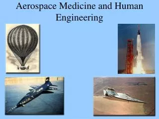
ENGINEERING AND MEDICINE
Dr. Mark H. Bechtel's story showcases the intersection of engineering and medicine. With a background in electrical engineering, inspired by family in healthcare, Bechtel pivoted to medicine in 1993. He navigated through medical school at the University of Washington and completed residency in radiology at the University of Wisconsin. His expertise led him to become a musculoskeletal specialist, emphasizing the critical role engineers play in medical fields, particularly in radiology, where technology is paramount. Bechtel illustrates how engineering principles enhance medical practice, bridging the gap between technical proficiency and patient care.

ENGINEERING AND MEDICINE
E N D
Presentation Transcript
ENGINEERING AND MEDICINE BY Mark H. Bechtel, M.D.
My Story • No inclination into medicine originally • Mother and wife are nurses, Wife also CRNA • 1st hand experience with hospitalization • Wanted Change • Career Counseling • Decided on Medicine in 1993 • Prerequisites by 1994 and started Med School.
Introduction • Moscow High School • BSEE, University of Idaho 1989 • Internships at Varian and Chevron during EE training. • Test Engineering at IBM, 1989-1991 • VLSI Design at AHA, 1991-1994 • MD at University of Washington 1994-1998 • Internship in Spokane, Washington 1999 • Radiology Residency at University of Wisconsin, 1999-2003 • General Radiologist in Brainerd, MN 2003-2004 • Musculoskeletal Fellowship at Penn State Hershey, 2004 • General Radiologist, MSK Specialist, Yankton, SD, 2005-2006 • General Radiologist, MSK Specialist, Moscow, ID, 2006-present
Main Points • Engineers as Physicians • Engineers as Information System Experts • Biomedical Engineering • Electrical Design in Medicine
Physician Engineers • Engineering is an excellent base for medicine • High percentage of radiologists are engineers • Engineering teaches a method of thinking that is not taught in other undergrad degrees
Medical School • 4 Years • Easier conceptually than engineering • More time than engineering • Engineering: if understand the concept then studying is over. • Medicine: Doesn’t matter if understand the concept. Human body is dynamic and the patient is still sick. Learning is constant and there is no definite endpoint. • Much more memorization
Internship • Most Physicians have internships • One year general training • Interview and selection process
Residency • Three to Six years • Radiology (4 years)
Fellowship • Further specialization • 1-2 years for radiology • …
Engineers as Information System Specialist • Radiology is highly Technology Dependent • PACS systems • Large storage system • Single CT can have 2000 images at 500Kbyte each • Need to interface with different equipment • Need to be able to send entire studies many miles away • NightHawk
Biomedical Engineering • Designing equipment for medical use • Ie: Insulin pump and detector • Pacemaker/defibrillator • Digital Subtraction Angiography • Stents • Intravascular work
Pacemaker • Earl Bakken
Pacemaker • Bakken’s orignal schematic
Pacemaker • Newer Devices
Pacemaker • Chest Xray
Pacemaker • Conduction system
MRI Images • Enhancement characteristics
MRI Images • Diffusion Tensor Imaging
MRI Images • MRA
MRI Images • Fat saturation
MRI Images • Spectroscopy
MRI Images • Cardiac Imaging
MRI Images • K-Space
CT • See other lecture
Conclusion • Engineering is an excellent base for medicine as a researcher, designer, information specialist, or as a physician.
Multidetector CT Mark Bechtel, M.D.
Education • Medical School: University of Washington • Radiology Residency: University of Wisconsin • Musculoskeletal Fellowship: Penn State University, Milton S. Hershey Medical Center
Chronological Developments in Multisclice CT • 1971 CT invented by Godfrey Hounsfeld of EMI and independently by Allan Cormack of Tufts University, Massachusetts. • 1974-1976 First Commercial CT scanners (for head CT only) • 1976 Whole body CT now available. • 1980 CT now widely available. • 1989 Introduction of Helical CT by Siemens, Germany • 1991 Launch of Dual Slice CT by Elscint, Haifa, Israel • 1999 Launch of 4 Slice Scanners • 2002 Launch of 16 Slice Scanners • 2003 Prototype 32 Slicers developed • 2003 Prototype 256 Slicers developed (Toshiba) 4D CT • 2003 Research in Flat Panet Detectors • 2003 Research in Faster scanning (<0.4 s rotation time) • 2003 Research in Cone Beam CT Multislice CT : A Quantum Leap in Whole Body ImagingIK indrajit, mn shreeram, jd d’souzaInd J Radiol Imag 2004 14:2:209-216
16 Slice is new standard • 32 and 64 slice models for cardiac scanning • New method of use is 3D evaluation versus axial imaging
Evaluation of a Mandibular Lesion • Left mandibular lesion was scanned in the axial and coronal planes. • Sagittal, oblique Sagittal and 3D images were reformated.
Comparison of Reconstructions • Comparing lumbar spine reconstructions from usual abdominal CT data sets from a single slice CT scanner and from a 16 slice multidetector CT.
3D Reconstruction of Bones and Fractures • Multiplanar reconstructions are possible • Allows better visualization of orientation of certain types of fractures. • Experienced readers often prefer 2D reconstructions
CTA of the Lower Extremities • Fast scanning abilities allows scanning of the lower extremities for vascular disease. • Makes conventional diagnostic angiography almost obsolete. • Can be used for surgical planning.